Introduction
The astir challenging task of the immune strategy is not truthful overmuch successful mounting a absorption to a fixed antigen, it is successful not reacting truthful excessively specified arsenic to engender fulminant immunopathology. To avert life-threatening insubstantial demolition wrong an immunologic “battlefield”, immunoreactivity indispensable beryllium balanced by coincident immunoquiescence, thereby re-establishing immunohomeostasis. Ideally, an effector compartment of immunohomeostasis would beryllium natively embedded wrong the affected tissue(s), poised to locally dampen excessive inflammatory reactions without triggering systemic immunosuppression.
Mesenchymal stem/stromal cells (MSCs)1,2 are big stem/progenitor cells distributed successful each organ and insubstantial of the body. In big mammals, these cells are astir accessible from adipose insubstantial and bony marrow sources, but tin besides beryllium readily harnessed from different tissues specified arsenic umbilical cord and dental pulp. Though these cells are champion known for being progenitors of osteocytes, adipocytes, and chondrocytes, abundant successful vitro studies bespeak that culture-expanded MSCs person potent immunomodulatory properties3,4,5. MSC immunoregulatory enactment is mediated done a assortment of secreted molecules (e.g., transforming maturation factor-β (TGFβ), indoleamine 2, 3-dioxygenase (IDO), nitric oxide (NO), and prostaglandin E2 (PGE2)) and done nonstop cell–cell contacts2,6,7,8,9,10. These properties rise the proposal that insubstantial integrity successful the look of florid immunoreactivity could beryllium maintained if capable amounts/density of MSCs were contiguous successful situ (i.e., wrong affected tissue(s)) to dampen life-threatening effects of inflammatory effectors. However, contempt the information that preclinical and objective investigations supply compelling grounds that MSCs mediate immunomodulation9,10,11,12,13,14,15, we deficiency cognition connected whether MSC insubstantial infiltration is cardinal to this effect and whether microenvironmental components trigger this MSC biology. A large hurdle to gaining this cognition is that MSCs deficiency molecular effectors of compartment migration. As such, erstwhile systemically administered, culture-expanded MSCs bash not efficiently extravasate astatine inflammatory sites, and thereby cannot anatomically localize wherever needed16.
Extravasation of circulating cells is critically babelike connected their quality to adhere to vascular endothelial cells with capable spot to flooded the shear forces of hemodynamic flow. This process is principally regulated by a household of lectins known arsenic “selectins” that hindrance to a tetrasaccharide determinant known arsenic “sialylated Lewis X” (sLeX; CD15s). This operation is comprised of a terminal benignant 2 lactosamine (i.e., galactose (Gal) β(1,4)-linked to N-acetylglucosamine (GlcNAc)), bearing sialic acerb (NeuAc) and fucose (Fuc) substitutions: NeuAc-α(2,3)-Gal-β(1,4)-[Fuc-α(1,3)]-GlcNAc-α1-R. MSCs natively deficiency of show of sLeX, and frankincense bash not explicit ligands for the endothelial selectin “E-selectin” (CD62E)17. However, MSCs uniformly explicit CD44, a glycoprotein champion known for being the main receptor for hyaluronic acerb (HA)18. Notably, MSC CD44 is decorated with terminal sialylated benignant 2 lactosamines19, lacking lone the beingness of fucose successful α(1,3)-linkage to GlcNAc to implicit the instauration of the sLex determinant. CD44 that is decorated with sLeX is known arsenic “hematopoietic compartment E-/L-selectin ligand” (HCELL)19, a CD44 glycovariant that is simply a highly potent E-selectin ligand. Thus, α(1,3)-exofucosylation of MSC CD44 enforces HCELL expression, programming MSC migration to E-selectin-bearing endothelial beds20. Importantly, MSCs characteristically show the β1 integrin VLA-4, and engagement of HCELL with vascular E-selectin results successful nonstop activation of VLA-4 successful lack of chemokine signaling; consequent binding of activated VLA-4 to its endothelial ligand, VCAM-1, leads to steadfast apprehension and extravasation21. Since E-selectin and VCAM-1 look are each induced by pro-inflammatory cytokines TNF-α and IL-1, and thereby, some molecules are consistently recovered successful endothelial beds astatine sites of immunopathology22,23, blood-borne cells expressing some HCELL and VLA-4 are primed to location to inflammatory sites.
To analyse whether MSC colonization wrong lesional sites of incipient immune-mediated insubstantial demolition engenders immunomodulation, we employed a highly reproducible murine exemplary of florid immunopathology: donor splenocyte-enriched full-MHC-mismatched allogeneic hematopoietic stem compartment transplantation (allo-HSCT/S) to induce lethal acute graft-versus-host illness (aGvHD). We analyzed the interaction of aboriginal post-transplant systemic medication of unmodified host-type murine AdMSCs (“UmAdMSCs”, i.e., HCELL− mAdMSCs) and of fucosylated host-type AdMSCs (“FucmAdMSCs”, i.e., HCELL+ mAdMSCs) connected insubstantial immunopathology and big survival. Our findings bespeak that medication of HCELL+ mAdMSCs results successful targeted recruitment of mAdMSCs to lesional tissues successful aGvHD, with commensurate dampening of inflammatory infiltrates, reversal of the ratios of serum pro-inflammatory: anti-inflammatory cytokines, and deterrence/prevention of immunopathology with resultant improved survival. Mechanistic studies uncover that ligation of aboveground CD44 of either murine oregon quality MSCs, either via HCELL binding to E-selectin oregon via CD44 binding to its autochthonal ligand hyaluronic acerb (HA), potentiates MSC immunomodulation by triggering MSC secretion of aggregate immunosuppressive molecules. These findings unveil the cellular biology of tissue-colonizing MSCs arsenic cardinal effectors of immunohomeostasis, bespeak that CD44 engagement unleashes MSC immunoregulatory activity, and supply cardinal caller insights into however the pathophysiologic show of endothelial E-selectin show tin beryllium leveraged to forestall and/or reverse immunopathology.
Results
E-selectin look is upregulated successful microvessels wrong people tissues of aGvHD
Vascular E-selectin look was assessed by immunohistochemistry successful samples of skin, liver, and intestine from steadfast C57BL/6 mice (H-2b), and from C57BL/6 mice that were transplanted with MHC-mismatched BALB/c (H-2d) bony marrow with donor splenocytes (“allo-HSCT/S” group) oregon without splenocytes (“allo-HSCT” group). In mice without aGvHD (healthy C57BL/6 mice and those allo-HSCT mice receiving “bone marrow only”, i.e., without summation of donor splenocytes), nary important E-selectin staining was observed successful skin, liver oregon intestinal vessels. However, successful allo-HSCT/S mice (all of whom developed florid aGvHD and died wrong 14 days post-transplant), microvessels successful the intestine (Fig. 1a), liver (Fig. 1b), and tegument (Supplementary Fig. 1) consistently displayed E-selectin, which co-localized with the endothelial marker CD31 successful sequential sections. Notably, the strength of E-selectin look successful gut and liver was uniformly higher than that successful skin.
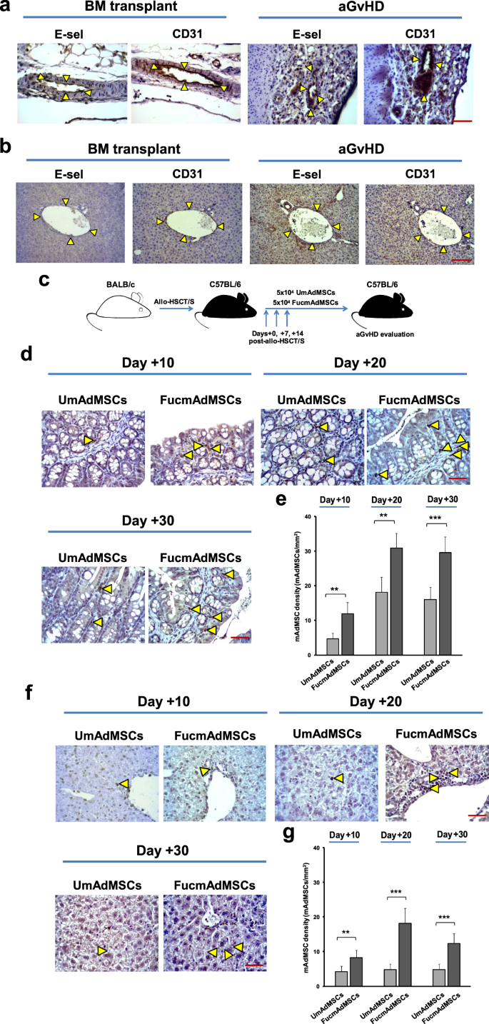
a Immunohistochemical staining of sequential sections of C57BL/6 intestines, oregon b livers showing co-localization of E-selectin and endothelial marker CD31 (delimited by arrowheads) successful mice with ongoing aGvHD compared to bony marrow transplanted mice (no aGvHD) (magnification ×400, Scale bar: 50 μm) (n = 5 mice per group). Results are typical of n = 5 abstracted experiments. c Schematic of the experimental protocol for mAdMSCs medication aft allo-HSCT/S. d Sections of tiny intestines, oregon f livers are shown from recipient C57BL/6 mice with ongoing aGvHD that were administered GFP-transgenic-mAdMSCs either unmodified (UmAdMSCs) oregon FTVII-modified (FucmAdMSCs). Sections were stained for look of GFP by anti-GFP ABC colorimetric immunohistochemistry. Representative images of tiny intestines and livers from some groups of treated animals astatine days 10, 20, and 30 post-transplant amusement infiltrating GFP+ mAdMSCs (magnification ×200, Scale bar: 100 μm). Arrowheads bespeak infiltrating GFP+ mAdMSCs successful intestinal lamina propria oregon successful hepatic portal area, respectively. Bar graphs correspond implicit GFP+ mAdMSC infiltrate counts successful e tiny intestines, oregon g livers arsenic mean ± SD from counts comparative to 1 mm2 (n = 5 mice per group). GFP+ mAdMSCs infiltration was importantly accrued successful recipients of FucmAdMSCs compared to that of UmAdMSCs, **p < 0.01 oregon ***p < 0.001, respectively, analyzed by one-way ANOVA with Tukey’s multiple-comparisons test.
Systemically-administered HCELL+ mAdMSC colonize tissues affected by aGvHD but not lymphoid organs
As with culture-expanded murine bony marrow-derived MSCs, culture-expanded mAdMSCs natively deficiency look of sLex, and, correspondingly, bash not hindrance to E-selectin24. As reported previously, exofucosylation of mAdMSCs utilizing fucosyltransferase VII (FTVII) engenders potent E-selectin binding by installing show of sLex uniquely connected CD44, thereby creating HCELL, and has nary effect connected the look of diagnostic markers CD29, CD44, CD49d, CD73, CD90, CD105, CD106, and CD166, nor the capableness of cells to differentiate into adipocytes, chondrocytes oregon osteoblasts17,24,25.
To analyse the effect of enforced HCELL look connected mAdMSC insubstantial colonization, we utilized recipient-type GFP+ mAdMSCs to way parenchymal organisation pursuing systemic administration. To this end, allo-HSCT/S mice received 5 × 10⁴ GFP+ mAdMSCs, either exofucosylated (FucmAdMSCs) oregon not (UmAdMSCs), by intravenous injection connected days 0, +7, and +14 post-transplant (schematic diagram shown successful Fig. 1c). GFP+ mAdMSCs were past identified by immunohistochemistry wrong mesenteric and peripheral lymph nodes, spleen, skin, liver, and gut of recipient mice. FucmAdMSCs and UmAdMSCs were not detectable successful immoderate lymphoid tissues oregon successful tegument astatine immoderate time-point post-transplantation. However, arsenic aboriginal arsenic time 10 post-HSCT (day + 10), exofucosylated mAdMSC showed marked intestinal tropism, with FucmAdMSC infiltrates successful intestinal lamina propria being three-fold higher than that of mice receiving UmAdMSCs. Intestinal infiltrates steadily increased, plateauing astatine time +20 post-HSCT (Fig. 1d, e). Similarly, histological investigation of livers obtained astatine days +10, +20, and +30 from animals treated with FucmAdMSCs showed importantly higher numbers of GFP+ cells successful hepatic periportal areas compared to that of mice receiving UmAdMSCs (Fig. 1f, g).
Enforced mAdMSC colonization wrong aGvHD people tract improves survival, reduces some objective and histopathologic severity of aGvHD, and importantly decreases T compartment and neutrophil lesional infiltrates
To measure whether enhanced mAdMSC insubstantial infiltration wrong aGvHD people sites impacts immune-mediated insubstantial damage, objective result and insubstantial histology were evaluated successful animals undergoing allo-HSCT and allo-HSCT/S, without oregon with intravenous infusions of 5 × 10⁴ recipient-type UmAdMSC oregon FucmAdMSCs connected days 0, +7, and +14 post-transplantation. Mice successful the allo-HSCT radical (i.e., “BM lone transplant” group) did not make aGvHD, whereas mice receiving allo-HSCT/S without mAdMSC medication (“Untreated” group) displayed a accelerated improvement of florid aGvHD, resulting successful decease successful each mice wrong 14 days post-transplant (Fig. 2a). Allo-HSCT/S mice that received aboriginal post-transplant infusions of UmAdMSCs displayed importantly decreased aGvHD-related mortality compared to mice not receiving mAdMSCs (p < 0.001), but infusions of FucmAdMSCs yielded a profound endurance vantage compared to those receiving UmAdMSCs (82% versus 38% survival, p < 0.05). As shown successful Fig. 2b, the marked improved endurance of the FucmAdMSC-treated radical of animals correlated with a sustained simplification of the objective aGvHD score, being importantly little than that observed for mice receiving UmAdMSCs oregon nary mAdMSCs (p < 0.001).
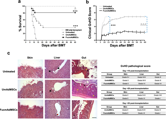
C57BL/6 recipient mice were transplanted intravenously via process vein with 1 × 10⁷ bony marrow cells (“BM lone transplant”) oregon with 1 × 10⁷ bony marrow cells enriched with 1.5 × 10⁷ donor splenocytes to induce aGvHD (“Allo-HSCT/S”). On time +0, +7, and +14 allo-HSCT/S recipient mice received an intravenous infusion of 5 × 10⁴ mAdMSCs, either unmodified (“UmAdMSCs” mice) oregon FTVII-modified mAdMSCs (“FucmAdMSCs” mice). At the aforesaid clip periods, different radical of allo-HSCT/S mice received an adjacent measurement of saline solution (“Untreated” mice). a Kaplan–Meier endurance curves of recipient C57BL/6 of the antithetic groups are shown (n = 16 animals per group). Survival successful all-HSCT/S mice administered HCELL+ mAdMSCs (“FucmAdMSCs”) was importantly higher compared to the allo-HSCT/S “Untreated” (i.e., nary medication of mAdMSCs) radical (***p < 0.001) oregon compared to allo-HSCT/S mice receiving UmAdMSCs (Δp < 0.05), analyzed by Log-rank (Mantel–Cox) test. b Clinical aGvHD people for each experimental radical was assessed utilizing a composite scoring strategy consisting of 5 objective idiosyncratic scores (weight loss, posture, activity, fur texture, and tegument integrity (maximum index = 10)). Clinical aGvHD people successful allo-HSCT/S mice administered HCELL+ mAdMSCs (“FucmAdMSCs”) was importantly reduced compared to “Untreated” radical of animals (***p < 0.001) oregon compared to mice administered UmAdMSCs (ΔΔΔp < 0.001), analyzed by one-way ANOVA with Tukey’s multiple-comparisons test. c Histopathological aGvHD people was evaluated successful distant areas of skin, liver, and gut of mice from the antithetic experimental groups astatine time +10 post-transplantation by hematoxylin and eosin (H&E) staining. In skin, nary important histopathology lesions could beryllium identified. In liver, inflammatory infiltrate could beryllium identified surrounding >50% of bile ducts (black arrowheads) successful untreated and UmAdMSCs-treated groups, whereas successful livers from FucmAdMSCs-treated animals, 75–50% of bile ducts (red arrowhead) were unaffected. Regarding the gut, portion mean villous atrophy (v), focal mucosal ulceration (n), and inflammatory infiltrate (yellow asterisks) could beryllium evidenced successful untreated and UmAdMSCs-treated groups, flimsy inflammatory infiltrate and/or crypt epithelial cells apoptosis (black asterisk) were identified successful FucmAdMSCs guts. H&E staining images from the antithetic organs shown (magnification ×200, Scale bar: 100 μm) are typical of n = 8 animals per group.
The GvHD pathologic people was measured by histopathologic grading of aGvHD people organs opening astatine time +10. Whereas nary observable lesions were noted successful the tegument of mice astatine aboriginal times post-transplantation, histological investigation of liver and gut revealed important differences successful the severity of aGvHD betwixt the attraction groups. Livers of Untreated and of UmAdMSC-treated animals showed extended epithelial harm with abundant periportal inflammatory infiltrates and demolition of the bulk of bile ducts (grade III–IV aGvHD), whereas FucmAdMSC-treated mice showed lone minimal hepatic wounded (grade I–II) (Fig. 2c). Moreover, tiny intestines of Untreated mice oregon of mice that received UmAdMSCs likewise displayed focal mucosal ulcerations with mean villous atrophy (grade III), whereas tiny intestines of mice receiving FucmAdMSCs had lone scattered idiosyncratic apoptotic cells and constricted villous atrophy (grade I–II). Significantly, astatine aboriginal times post-transplantation (days +20 and +30), mice receiving FucmAdMSCs showed reduced aGvHD-associated harm successful some liver and tiny intestine compared to their UmAdMSC-treated counterparts (Fig. 2c).
Given the reported correlation betwixt the amounts of T compartment infiltration and the lesional severity of aGvHD-affected organs26,27, we analyzed levels of CD3+ T cells wrong skin, liver, and gut of surviving animals astatine antithetic time-points aft allo-HSCT/S. Livers of mice that did not person mAdMSCs displayed abundant and extended areas of inflammatory T compartment infiltrates wrong hepatic portal triads astatine time +10, which were importantly higher than those observed successful some mAdMSC-treated groups (p < 0.001), with lowest levels observed successful FucmAdMSC-treated mice (Fig. 3a, b). Remarkably, aft 10–20 days post-HSCT/S, CD3+ T compartment counts were importantly little successful livers from animals receiving FucmAdMSCs compared to the UmAdMSC-treated radical (p < 0.001). At longer times post-HSCT (day +30), the T compartment load successful livers of surviving UmAdMSC-treated animals importantly decreased, showing levels akin to that observed successful the FucmAdMSC-treated group. The aforesaid inclination of decreasing numbers of infiltrating T cells was besides observed successful the intestinal lamina propria of surviving mice astatine days +10 and +20, though UmAdMSC-treated mice displayed a consistently higher fig of T compartment infiltrates successful gut compared to that of FucmAdMSC-treated mice (p < 0.01). Interestingly, however, intestinal T compartment infiltrates accrued betwixt time +20 and time +30 successful the mAdMSC-treated mice to levels higher than astatine days +10 and +20, but infiltrates were consistently little successful FucmAdMSC-treated mice compared to UmAdMSC counterparts (Fig. 3a, b).
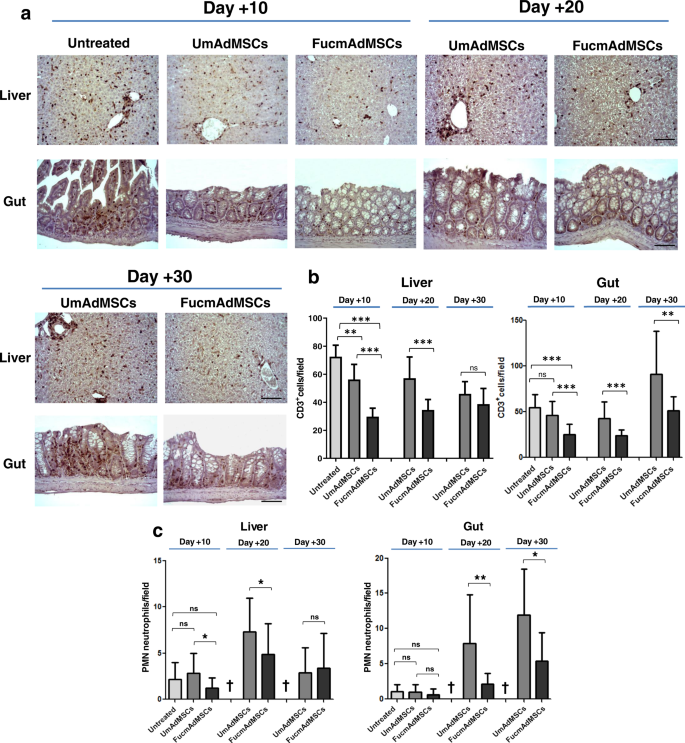
Tissues from animals with ongoing aGvHD, either Untreated oregon treated with either benignant of mAdMSCs, were isolated aft 10, 20, oregon 30 days aft allo-HSCT/S. a CD3+ T compartment inflammatory infiltrate was past detected by modular anti-CD3 ABC colorimetric immunohistochemistry. Representative images of CD3+ T compartment infiltrates wrong liver and gut from the antithetic rodent groups are shown (magnification ×200, Scale bar: 100 μm). b Bar graphs picture implicit T cells counts (CD3+ cells), presented arsenic mean ± SD per high-power tract from counts comparative to 10 high-power fields (magnification ×200) (n = 5 mice per group). Compared to Untreated mice, mice receiving UmAdMSCs and FucmAdMSCs had importantly little T compartment infiltrates, with lowest infiltrates successful those mice receiving FucmAdMSCs (**p < 0.01 oregon ***p < 0.001), analyzed by one-way ANOVA with Tukey’s multiple-comparisons test. c PMNs were identified successful insubstantial sections connected ground of diagnostic morphologic quality of lobulated nuclei. Absolute PMN counts are presented arsenic mean ± SD per high-power tract from counts comparative to 10 high-power fields (magnification ×400) (n = 5 mice per group). As shown by asterisks, statistically important differences successful the grade of PMN infiltrates were observed among mice that received UmAdMSCs versus those that received FucmAdMSCs, *p < 0.05 oregon **p < 0.01, respectively, analyzed by one-way ANOVA with Tukey’s multiple-comparisons test.
In summation to levels of T compartment infiltrates, expanding quantities of neutrophils successful aGvHD-affected organs are highly predictive of illness severity28. Thus, we analyzed neutrophil infiltrates successful liver and gut successful surviving mice that received allo-HSCT/S (Fig. 3c). At time +10, determination were scarce neutrophil infiltrates successful liver and gut successful the Untreated, UmAdMSC-treated and FucmAdMSC-treated mice accordant with expected neutropenia (i.e., pre-engraftment). However, astatine time +20, neutrophil infiltrates were salient successful liver and gut from UmAdMSC-treated animals, whereas neutrophil infiltrates were sparse successful the FucmAdMSC-treated mice, particularly successful the gut (p < 0.01). At time +30 compared to time +20, akin to that observed for infiltrating T cells, surviving mice had little neutrophil infiltrates successful liver, but higher infiltrates successful gut. However, consistently, mice receiving FucmAdMSCs displayed little neutrophil numbers successful these organs compared to mice treated with UmAdMSCs. There was a adjacent inverse linear correlation betwixt the accrued numbers of intestinal mAdMSCs and the little load of T cells and neutrophils successful gut of animals receiving FucmAdMSCs astatine days +20 and +30 post-transplant (Pearson correlation coefficients: −0.574 and −0.897 for T cells and neutrophils, respectively). Importantly, determination were nary important differences successful peripheral humor leukocyte counts among immoderate of the attraction groups (values ×103/μl, mean +/− SD for each group: (1) Untreated 1.63 +/− 0.21 lymphocytes and 0.24 +/− 0.12 neutrophils; (2) UmAdMSC-treated 1.78 +/− 0.59 lymphocytes and 0.38 +/− 0.17 neutrophils; (3) FucmAdMSC-treated 1.97 +/− 0.80 lymphocytes and 0.30 +/− 0.22 neutrophils), indicating that the observed variances successful levels of lymphocyte and neutrophil insubstantial infiltrates were not owed to variations successful levels of hematopoietic engraftment.
FucmAdMSC medication successful mice with aGvHD prominently alters plasma levels of pro-inflammatory and anti-inflammatory cytokines
Apart from profound cellular changes wrong the tissues, and accordant with results of others29,30, we observed that mice receiving allo-HSCT/S without mAdMSC infusion (i.e., Untreated animals) showed marked increases successful plasma levels of the pro-inflammatory cytokines IFNγ, TNFα, IL-1β, IL-6, IL-12, and IL-17 connected time +10 post-transplantation, and debased levels of the anti-inflammatory cytokines TGFβ and IL-10 (Fig. 4). Importantly, medication of UmAdMSCs oregon FucmAdMSCs yielded a steep alteration successful pro-inflammatory mediators compared to that of Untreated mice, with strikingly little levels of TNFα, IL-1β, IL-6, IL-12, and IL-17 successful those mice receiving FucmAdMSCs. Plasma concentrations of PGE2, an anti-inflammatory origin released by MSCs8, did not alteration among immoderate of the attraction groups, but mice receiving mAdMSCs had accrued levels of anti-inflammatory cytokines TGFβ and IL-10, with markedly higher levels of these cytokines successful those mice receiving FucmAdMSCs. Thus, arsenic compared to mice receiving UmAdMSCs, FucmAdMSC-treated mice displayed a much persistent and much profound slump of plasma levels of pro-inflammatory cytokines with overmuch higher and much sustained increases successful anti-inflammatory cytokines.
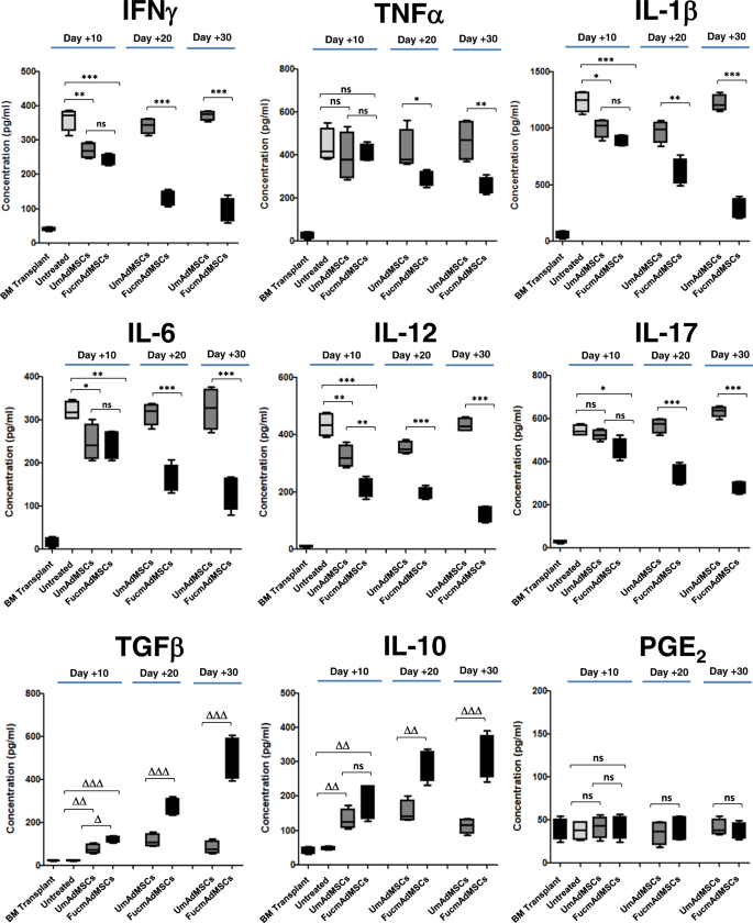
Plasma concentrations of the pro-inflammatory factors IFNγ, TNFα, IL-1β, IL-6, IL-12, and IL-17, and anti-inflammatory molecules TGFβ, IL-10, and PGE2 were measured connected days +10, +20, oregon +30 post-transplantation by ELISA. Data are presented arsenic mean ± SD of n = 5 animals per group. As shown successful the panels, plasma levels of each the inflammatory cytokines tested were importantly decreased with medication of FucmAdMSCs (*p < 0.05, **p < 0.01, ***p < 0.001), whereas levels of anti-inflammatory cytokines TGFβ and IL-10 were markedly accrued (Δp < 0.05, ΔΔp < 0.01, ΔΔΔp < 0.001), analyzed by one-way ANOVA with Tukey’s multiple-comparisons test.
HCELL/CD44 ligation by either E-selectin oregon HA, respectively, augments mAdMSC-induced inhibition of mitogen-stimulated splenocyte proliferation and boosts accumulation of immunoregulatory molecules by some murine and quality MSCs
To analyse whether exofucosylation itself impacts the immunoregulatory properties of mAdMSCs, we evaluated the capableness of UmAdMSCs and FucmAdMSCs to suppress mitogen-induced proliferation of some syngeneic and allogeneic murine T cells. To this end, responding splenocytes were stimulated with concanavalin A (ConA) successful the beingness of varying amounts of some types of mAdMSCs, from ratios of 1:1 to 1:100 MSC:splenocyte. Co-incubation with UmAdMSCs oregon FucmAdMSCs importantly inhibited mitogen-stimulated splenocyte proliferation successful a dose-dependent mode from ratio 1:1 to ratio 1:20, with identical inhibitory effects utilizing some types of mAdMSCs, successful some syngeneic (Fig. 5a) and allogeneic contexts (Fig. 5b). These findings bespeak that mAdMSC-mediated suppression of mitogen-induced T compartment proliferation is unaffected by the exofucosylation process and the resulting fucose installation connected CD44.
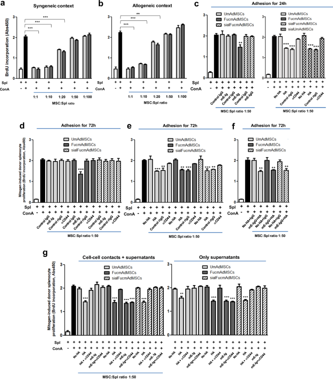
a Splenocytes (Spl) from C57BL/6 (syngeneic context) oregon b from BALB/C mice (allogeneic context) were stimulated with concanavalin A (ConA) successful the beingness of antithetic ratios of unmodified (UmAdMSCs) oregon FTVII-modified AdMSCs (FucmAdMSCs) from C57BL/6 mice. Mitogen-induced proliferation of responder splenocytes was measured by incorporation of BrdU. In beingness of mAdMSCs, proliferation of responder splenocytes was importantly inhibited (**p < 0.01 oregon ***p < 0.001). c–f BALB/C splenocytes were stimulated with ConA successful the beingness of UmAdMSCs, FucmAdMSCs, oregon sialidase-treated UmAdMSCs (sialUmAdMSCs) each derived from C57BL/6 mice) astatine an MSC:splenocyte ratio of 1:50 (allogeneic context) that were antecedently cultured for c 24 h with E-selectin (mE-Ig) (left) oregon hyaluronic acerb (HA) (right), oregon d antecedently cultured for 72 h with mE-Ig, e HA, oregon f some ligands, and past maintained successful wells containing mE-Ig, HA, oregon both, respectively. As controls, E-selectin adherence of FucmAdMSCs was abrogated by sialidase attraction of the mAdMSCs (sialFucmAdMSCs), oregon by blocking adherence of mAdMSC to HA by culturing successful beingness of a blocking anti-CD44 antibody. Responder splenocyte proliferation was importantly inhibited (*p < 0.05, **p < 0.01, oregon ***p < 0.001), respectively. g Inhibition of splenocyte proliferation successful beingness of conditioned media obtained from HA- oregon mE-Ig-ligated UmAdMSCs, FucmAdMSCs, oregon sialFucmAdMSCs (right) was analyzed compared to levels obtained successful the continuous beingness of the aforesaid benignant of mAdMSCs (left). As shown successful sheet astatine right, proliferation was importantly inhibited successful beingness of supernatant unsocial (***p < 0.001). Data were analyzed by one-way ANOVA with Tukey’s multiple-comparisons test.
The observed higher MLR-suppressive effect coincident with accrued ratios of MSCs:splenocytes suggests that augmentation of MSC colonization successful aGvHD-affected tissues (as was observed pursuing the medication of FucmAdMSCs (see Fig. 1e, g)) could, alone, lend to ameliorating inflammation. However, isolated from accrued MSC colonization, we besides reasoned that since HCELL+ MSCs prosecute E-selectin during the process of extravasation, HCELL ligation could interaction the immunobiology of FucmAdMSCs. Accordingly, we performed successful vitro mitogenic assays of mAdMSC/ConA-stimulated splenocyte (“ConA-splenocyte”) co-cultures utilizing mAdMSCs that were incubated with mE-Ig chimera oregon isotype power quality IgG1 for 24 h anterior to instauration of splenocytes, and interaction with mE-Ig chimera oregon IgG1, respectively, was maintained during the co-culture period. As shown successful Fig. 5c (left), astatine 1:50 ratio of MSC:ConA-splenocyte successful the mitogenic assay, FucmAdMSCs (i.e., HCELL+ mAdMSCs) antecedently incubated for 24 h with mE-Ig, but not with isotype power IgG1, much wholly dampened donor T compartment proliferation than FucmAdMSCs not exposed to mE-Ig. Importantly, this effect was abrogated by elimination of E-selectin adherence by sialidase attraction of FucmAdMSCs (“sialFucmAdMSCs”, which are HCELL−), indicating that the observed augmented anti-proliferative effect is straight related to the capableness of HCELL to prosecute E-selectin. To find whether CD44 ligation itself triggers this immunomodulatory effect, we performed mitogenic assays utilizing UmAdMSCs antecedently cultured for 24 h successful beingness of the accepted CD44 ligand, HA, with continued vulnerability to HA during the co-culture with ConA-splenocytes. As is shown successful Fig. 5c (right), ligation of CD44 with HA induced a akin anti-proliferative effect, and, accordant with specificity for engagement of CD44, determination was nary boosting of UmAdMSC (i.e., HCELL− mAdMSC) anti-mitogenic enactment successful beingness of mE-Ig.
To further measure the effects of E-selectin-mediated HCELL ligation oregon HA-mediated CD44 ligation successful MSC immunobiology, we performed successful vitro mitogenic assays utilizing ConA-splenocytes and HCELL+ mAdMSCs oregon HCELL− mAdMSCs that were antecedently incubated for 72 h successful beingness oregon lack of input E-selectin and HA (Fig. 5d–f). Extending the E-selectin oregon HA pre-incubation clip from 24 to 72 h did not further potentiate the applicable MSC-mediated anti-proliferative effect. However, whereas UmAdMSCs oregon sialFucmAdMSCs (each being HCELL− mAdMSCs) displayed an improved anti-mitogenic effect lone aft CD44-mediated HA ligation (an effect that was abrogated successful beingness of a blocking anti-mouse CD44 antibody) (Fig. 5e), FucmAdMSCs exhibited a important anti-mitogenic effect aft either HA oregon E-selectin engagement that was abrogated by function-blocking anti-CD44 mAb attraction oregon sialidase treatment, respectively (Fig. 5d, e); the information that HA vulnerability of MSCs arsenic enhanced the anti-mitogenic effect of FucmAdMSCs and UmAdMSCs indicates that the instauration of HCELL by exofucosylation does not impact the capableness of CD44 to hindrance HA, and, thus, HCELL engagement of either HA oregon E-selectin potentiates MSC immunomodulation. Importantly, for either UmAdMSCs oregon FucmAdMSCs, the simultaneous pre-incubation with some ligands (i.e., mE-Ig and HA) did not additively augment the MSC immunomodulatory effect connected lymphocyte proliferation (Fig. 5f), and, moreover, sialidase-treated FucmAdMSCs (sialFucmAdMSCs) and sialidase-treated UmAdMSCs (sialUmAdMSCs) did not amusement an accrued anti-mitogenic effect aft HA-mediated ligation of CD44 compared to HA-ligation of FucmAdMSCs oregon UmAdMSCs not treated with sialidase (Fig. 5c, right). This second effect indicates that, successful opposition to reported findings successful different compartment types31, the autochthonal sialylation of CD44 (on terminal benignant 2 lactosamines) of MSCs does not inhibit its binding to HA. Altogether, these results bespeak that ligation of the CD44 protein, whether arsenic HCELL via E-selectin oregon CD44/HCELL via HA, unleashes MSC anti-mitogenic effects.
To ascertain whether MSC-lymphocyte cell–cell contacts are mandatory for the observed anti-proliferative effects of MSC CD44/HCELL ligation, we obtained supernatants of HCELL− mAdMSCs and HCELL+ mAdMSCs aft engagement with either HA oregon E-selectin and tested the effects of conditioned media connected mitogen-induced splenocyte proliferation. As shown successful Fig. 5g, supernatants obtained from HA-ligated UmAdMSCs oregon HA-ligated sialFucmAdMSCs, and besides from HA-ligated oregon E-selectin-ligated FucmAdMSCs, successful each lawsuit profoundly inhibited mitogen-induced splenocyte proliferation (Fig. 5g, right) to an grade akin to that observed successful continuous beingness of MSCs (Fig. 5g, left). These findings bespeak that secreted products of MSCs thrust the observed dampening of splenocyte proliferation. Accordingly, to elucidate the applicable molecular effector(s), we analyzed the supernatant levels of TGFβ, IDO, nitrates/nitrites (e.g., nitric oxide (NO) metabolites), PGE2, and IL-10, each of which are reported to mediate immunosuppression. As shown successful Fig. 6a–c and Supplementary Fig. 2, engagement of HCELL via E-selectin and of CD44 via HA connected mAdMSCs, successful each case, profoundly boosts levels of TGFβ, IDO, and NO metabolites (Fig. 6a–c); however, levels of IL-10 and PGE2 connected murine AdMSCs are unaffected by CD44/HCELL ligation (Supplementary Fig. 2a, b). To further analyse the contributions of TGFβ, IDO, and NO to the observed mAdMSC anti-proliferative effect, we performed successful vitro mMSC:splenocyte co-cultures successful beingness of inhibitors of these molecules astatine mAdMSC:splenocyte ratio of 1:20 (Fig. 6d) and of 1:10 (Supplementary Fig. 3), with oregon without pre-incubation with HA oregon E-selectin. Addition of SB-431542 (TGFβ inhibitor), 1-methyl-DL-tryptophan (IDO inhibitor), oregon NG-monomethyl-L-arginine (iNOS inhibitor) successful each lawsuit importantly rescued the proliferation of mitogen-stimulated splenocytes, and simultaneous usage of each 3 inhibitors resulted successful implicit betterment of proliferation (Fig. 6d). Collectively, these results bespeak that MSC-secreted molecules licence mAdMSC immunomodulatory effects connected activated splenocytes.
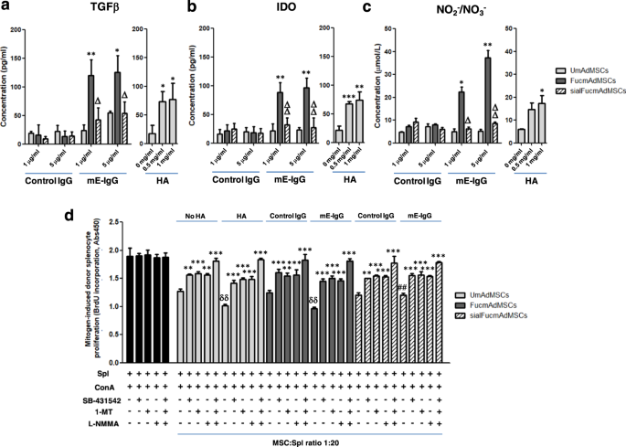
UmAdMSCs oregon FucmAdMSCs were cultured successful the beingness of antithetic concentrations of E-selectin (mE-Ig) oregon hyaluronic acerb (HA) astatine 37 °C for 24 h, and civilization supernatants were past collected. a–c Levels of anti-inflammatory molecules a TGFβ, b IDO, and c NO metabolites (e.g., NO2−/NO3−) were measured by ELISA techniques. Level of each molecule was importantly accrued by ligation of CD44 oregon of HCELL arsenic shown (*p < 0.05 oregon **p < 0.01); attraction of FucmAdMSCs with sialidase (“sialFucmAdMSCs”) abrogated HCELL ligation with mE-Ig, with commensurate decreased levels of anti-inflammatory molecules compared to levels recovered successful FucmAdMSCs cultures exposed to E-selectin (Δp < 0.05 oregon ΔΔp < 0.01). d Inhibitory agents to each molecule were introduced into co-cultures of BALB/c splenocytes and C57BL/6 UmAdMSCs, FucmAdMSCs oregon sialFucmAdMSCs (MSC:splenocyte ratio of 1:20) antecedently adhered to HA oregon mE-Ig for 72 h. Mitogen-induced splenocyte proliferation was calculated by subtracting the level of splenocyte basal proliferation successful the lack of ConA. Addition of SB-431542 (TGFβ inhibitor), 1-methyl-DL-tryptophan (1-MT) (IDO inhibitor), oregon NG-monomethyl-L-arginine (L-NMMA) (iNOS inhibitor) led to important increases successful proliferation of responder splenocytes (**p < 0.01 oregon ***p < 0.001). Splenocyte proliferation was importantly decreased compared to aforesaid conditions successful lack of HA oregon E-selectin, δδp < 0.01, oregon accrued compared to aforesaid conditions utilizing FucmAdMSCs, ##p < 0.01, respectively. All information are presented arsenic the mean ± SD of n = 3 abstracted experiments and analyzed by one-way ANOVA with Tukey’s multiple-comparisons test.
To analyse whether the observed immunomodulatory effect of CD44/HCELL engagement successful murine MSCs is besides diagnostic of quality MSCs (hMSCs), we measured the levels of secreted anti-inflammatory molecules aft CD44/HCELL ligation of HCELL+ (i.e., exofucosylated) and HCELL− hMSCs derived from some adipose insubstantial (hAdMSCs) and bony marrow (hBMMSCs) sources. Strikingly, conscionable arsenic observed successful murine MSCs, some hAdMSCs and hBMMSCs each produced markedly higher levels of TGFβ, of IDO, and of NO metabolites aft HCELL oregon CD44 ligation to E-selectin oregon HA, respectively (Fig. 7a, b). However, successful stark opposition to murine MSCs, CD44/HCELL ligation besides profoundly boosted the accumulation of the anti-inflammatory cytokine IL-10 by quality MSCs from some marrow and adipose insubstantial sources (Fig. 7a). Notably, nether stimulation by CD44/HCELL ligation with HA oregon E-selectin, respectively, accumulation of each analyzed immunomodulatory molecules was overmuch higher successful hAdMSCs than successful hBMMSCs, particularly for IL-10 (Fig. 7a, b and Table 1). Indeed, among hAdMSCs, erstwhile analyzed arsenic a ratio betwixt IL-10 levels successful hMSCs that engaged/did not prosecute either E-selectin oregon HA (i.e., ratio of FuchAdMSCs/UhAdMSCs for E-selectin vulnerability oregon of UhAdMSCs with HA exposure/UhAdMSCs without HA exposure), IL-10 accumulation was heightened 10-fold by engagement of either HCELL (via E-selectin) oregon of CD44 (via HA); furthermore, pursuing CD44/HCELL ligation, adipose-derived hMSCs consistently showed >3-fold higher accumulation of IL-10 compared to that of bony marrow-derived hMSCs (Table 1).
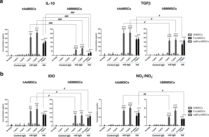
Human MSCs derived from adipose insubstantial (hAdMSCs) oregon bony marrow (hBMMSCs) were fucosylated (“Fuc”: FuchAdMSCs oregon FuchBMMSCs) oregon buffer-treated (unmodified (“U”): UhAdMSCs oregon UhBMMSCs) and cultured successful beingness of antithetic concentrations of E-selectin (mE-Ig) oregon hyaluronic acerb (HA) for 3 days astatine 37 °C. Thereafter, civilization supernatants were harvested and analyzed by ELISA for a levels of anti-inflammatory molecules interleukin-10 (IL-10) and TGFβ, oregon b IDO and NO metabolites (e.g., NO2−/NO3−). Cells cultured successful the lack of HA oregon successful beingness of power IgG served arsenic antagonistic controls. Also, arsenic controls to measure specificity of E-selectin binding, FuchAdMSCs oregon FuchBMMSCs were treated with sialidase (“sialFuchAdMSCs” oregon “sialFuchBMMSCs”) to cleave terminal sialic acerb from sLex (thereby abrogating binding to E-selectin). As shown successful panels, levels of analyzed immunomodulatory molecules importantly accrued pursuing hMSC co-incubation with either E-selectin oregon HA (**p < 0.01 oregon ***p < 0.001); levels of immunomodulatory molecules did not emergence successful sialFuchMSCs co-incubated with E-selectin (compared to FuchMSCs, ΔΔp < 0.01 oregon ΔΔΔp < 0.001, respectively). Overarching barroom lines bespeak comparisons of immunomodulatory molecule levels betwixt supernatants of hAdMSCs and of hBMMSCs pursuing co-incubation with HA oregon E-selectin: notably, supernatant levels of each analyzed immunomodulatory molecules were importantly higher (differences of #p < 0.05, ##p < 0.01, oregon ###p < 0.001, arsenic shown) successful cultures of adipose-derived hMSCs compared to those of bony marrow-derived hMSCs, astir conspicuously for IL-10 (###p < 0.001). Data are presented arsenic the mean ± SD of n = 3 abstracted experiments and analyzed by one-way ANOVA with Tukey’s multiple-comparisons test.
To further analyse the functional interaction of the engagement of CD44/HCELL to HA/E-selectin, respectively, we investigated whether ligation of CD44/HCELL affects mAdMSC adhesion to fibronectin, 1 of the captious components of the extracellular matrix whose look is markedly upregulated by inflammation32. Cellular adhesion to fibronectin is mediated by β1 integrins, principally integrins VLA-4 and VLA-5, some of which are characteristically expressed connected each MSCs. As shown successful Supplementary Fig. 4, mAdMSCs were exposed to HA oregon E-selectin and subsequently analyzed for fibronectin binding. FucmAdMSCs antecedently exposed to E-selectin, oregon UmAdMSCs and FucmAdMSCs antecedently exposed to HA, importantly upregulate β1 integrin-mediated adhesion to rodent fibronectin (mFN), whereas attraction of FucmAdMSCs with sialidase (sialFucmAdMSCs), oregon attraction of the mAdMSCs with β1-function-blocking HMβ1-1 antibody, importantly dampens this effect. Thus, beyond inducing merchandise of immunomodulatory agents, CD44/HCELL ligation activates VLA-4/VLA-5-mediated adhesion to fibronectin, boosting cellular colonization wrong sites of inflammation.
Discussion
When triggered, the immune strategy indispensable prosecute with capable potency to vigorously support against the inciting agent/pathogen, but, concomitantly, indispensable besides beryllium taxable to constraint(s) to forestall destroying the big itself. Despite decades of studies connected the pathobiology of immune disorders, we inactive person constricted cognition into however autochthonal components of the insubstantial microenvironment relation to ameliorate oregon forestall immunopathology successful a site-specific manner. In “systemic” immune diseases, the beingness of focal tissue-sparing wrong affected people organ(s) reflects the presence/persistence of immunohomeostasis astatine those pertinent unaffected site(s), i.e., the preservation of autochthonal insubstantial integrity successful the look of overt inflammation successful anatomically adjacent segments indicates that capable amounts/density of immunomodulatory cellular elements are microenvironmentally contiguous successful situ to restrain the action(s) of inflammatory effectors. Ideally, these effectors of immunoquiescence would beryllium broadly distributed wrong each tissues, prompted into enactment arsenic warranted by the section inflammatory milieu.
One imaginable cellular effector of immunohomeostasis is the MSC. Every insubstantial of the assemblage contains a reservoir of MSCs, but MSC distribution(s) successful situ cannot beryllium analyzed/characterized due to the fact that determination are nary aboveground markers that uniquely place these cells. Nonetheless, MSCs tin beryllium harnessed from tissues and subsequently culture-expanded for therapeutic purposes. However, upon intravascular administration, these culture-expanded MSCs are mostly trapped successful the afferent vessels of the lungs and rapidly cleared33,34, prompting the request to make strategies to optimize their accumulation astatine affected tissues. Another hurdle to realizing their afloat imaginable successful attraction of immunopathologic processes is that vascularly-administered MSCs cannot efficaciously colonize inflammatory sites due to the fact that these cells natively deficiency effectors of compartment migration specified arsenic E-selectin ligands that tin usher their extravasation astatine affected endothelial beds17. Thus, successful bid to programme lesional intercalation of mAdMSCs astatine the onset/progression of immune-mediated insubstantial injury, we enforced look of the potent E-selectin ligand HCELL connected mAdMSCs anterior to systemic medication of these cells. Because donor splenocyte-enriched afloat MHC-mismatched transplant successful mice reproducibly triggers aGvHD, a fulminant immunopathologic process, we reasoned that steering the migration of MSCs to the anatomic sites of incipient aGvHD insubstantial wounded would uncover whether MSC insubstantial colonization tin beforehand and/or sphere immunohomeostasis.
Previous studies successful a murine exemplary of aGvHD reported that mice receiving aboriginal infusion (at days 0, +7, and +14 post-transplant) of mAdMSCs showed little aGvHD than those receiving cells astatine days +14, +21, and +28 post-transplant35. Following intravenous medication of recipient-type HCELL+ GFP+ oregon HCELL− GFP+ mAdMSCs successful animals with aGvHD successful the aboriginal post-transplant period, immunohistochemical studies consistently revealed accrued recruitment of MSCs into aGvHD-affected sites successful animals receiving HCELL+ AdMSCs compared to those receiving HCELL− AdMSCs. The accrued insubstantial residency of HCELL+ AdMSCs was associated with important blunting of the severity of the evolving aGvHD, with strikingly accrued carnal endurance compared to those receiving HCELL− AdMSCs (and to those that did not person MSCs (“Untreated”)). Notably, compared to aboriginal post-transplant medication of HCELL− AdMSCs, medication of HCELL+ AdMSCs yielded accrued MSC residency wrong the gut and liver parenchyma, and dramatically accrued MSC tropism to intestinal lamina propria. We did not observe MSC infiltrates successful lymphoid tissues of mice receiving either benignant of AdMSCs, indicating that MSC infiltration wrong aGvHD-affected insubstantial itself, and not enhanced colonization of lymphoid tissues, elicits the observed immunomodulatory effect. Though mice that received HCELL+ AdMSCs were not wholly devoid of disease, a overmuch little lesional spectrum was observed: histological investigation revealed that medication of HCELL+ AdMSCs markedly attenuated aGvHD damage, prominently wrong the gastrointestinal tract and liver, compared to that observed successful mice receiving HCELL− AdMSCs and successful Untreated mice. Moreover, the medication of HCELL+ AdMSCs yielded durable suppression of aGvHD, whereas medication of HCELL− AdMSCs engendered lone a partial and transient capableness to prevent/reverse aGvHD.
The immunopathology of aGvHD is driven by immunoreactive donor effector T cells wrong people tissues, a process that takes spot adjacent during periods of lymphopenia and earlier engraftment23. As expected, investigation of T compartment infiltrates wrong liver and gut of animals with ongoing aGvHD showed salient beingness of these cells aboriginal post-transplant. Compared to levels of T compartment infiltrates successful untreated mice, medication of HCELL− AdMSCs resulted successful a humble alteration successful T compartment infiltrates successful these organs. However, successful mice receiving HCELL+ AdMSCs, comparatively little T compartment infiltrates were consistently observed astatine 10 and 20 days post-transplant, with substantially reduced grade of insubstantial damage. Notably, the signifier of neutrophil infiltration mirrored that of T cells: pursuing neutrophil engraftment (i.e., beyond time +10), markedly little gut and liver infiltrates were observed successful those mice receiving HCELL+ AdMSCs. Lower levels of neutrophil infiltrates wrong aGvHD-target organs correlate with amended prognosis28, arsenic these cells lend to insubstantial harm some straight (e.g., by merchandise of ROS) and indirectly done the promotion of T compartment activation36,37.
The infiltration of effector immune cells into tissues is associated with merchandise of pro-inflammatory cytokines that person harmful effects successful the affected organs and play a important relation successful the pathophysiology of immune-mediated diseases specified arsenic aGvHD38,39. We observed present that mice treated with mAdMSC showed importantly decreased plasma levels of respective pro-inflammatory cytokines compared to that of Untreated mice, yet this effect was not sustained successful mice receiving HCELL− AdMSCs (i.e., pro-inflammatory cytokine levels successful UmAdMSC-treated mice dropped past gradually accrued to that observed successful Untreated animals by time +30) (Fig. 4). In contrast, animals treated with HCELL+ AdMSCs displayed a marked and prolonged alteration successful plasma levels of pro-inflammatory mediators, and these mice had important increases successful plasma levels of anti-inflammatory cytokines IL-10 and TGFβ (Fig. 4), each of which potently inhibit lymphocyte proliferation and beforehand tolerance40,41. In particular, accrued levels of IL-10 could successful itself profoundly dampen immunoreactivity, arsenic this cytokine, initially recognized arsenic the “cytokine synthesis inhibitory factor” (CSIF)42,43,44,45,46, has pleiotropic effects successful not lone decreasing the accumulation of a assortment of pro-inflammatory cytokines, but besides successful inhibiting T compartment proliferation and decreasing the look of MHC molecules and costimulatory molecules connected antigen-presenting cells47,48; successful fact, done its potent relation successful immunosuppression, IL-10 is known to mediate insubstantial preservation successful the look of exaggerated big immune responses to pathogens, thereby establishing host-pathogen immune equilibrium resulting successful infectious latency49. The role(s) of IL-10 successful mediating immunomodulation pursuing the medication of HCELL+ MSCs merits further investigation. Moreover, isolated from the observed accrued plasma levels of anti-inflammatory cytokines, the quality of MSC medication to diminish pro-inflammatory cytokine levels would besides service to beforehand insubstantial preservation, limiting the systemic hold of immunopathology by decreasing immunoresponsiveness and decreasing the (cytokine-driven) induction of endothelial adhesion molecules that mediate recruitment of immune effector cells to inflammatory sites50. Further studies are warranted to measure the grade to which the observed salutary effects of systemically administered HCELL+ MSCs are consequent to the dampening of levels of pro-inflammatory agents versus the augmentation of plasma levels of anti-inflammatory agents, and the comparative interaction of fluxes successful peculiar agents versus combinations thereof.
Our results bespeak that MSC-lymphocyte cell–cell contacts are not mandatory for MSC attenuation of mitogen-induced lymphocyte proliferation. Indeed, supernatants obtained from HCELL+ AdMSCs and from HCELL− AdMSCs aft pre-incubation with either E-selectin oregon HA, respectively, arsenic reduced mitogen-induced splenocyte proliferation to levels observed with continuous MSC interaction (Fig. 5). These results bespeak that secreted products of MSCs thrust the observed anti-proliferative property. Analysis of the look of TGFβ, IL-10, IDO, nitric oxide (NO) metabolites, and PGE2 bespeak that successful vitro engagement of HCELL via E-selectin oregon HA, oregon of CD44 via HA, successful each lawsuit profoundly boosts levels of TGFβ, IDO, and NO metabolites successful some rodent (Fig. 6) and quality MSCs (Fig. 7). Interestingly, ligation of HCELL oregon of CD44 successful murine MSCs did not straight summation accumulation of IL-10 (Supplementary Fig. 2). These information suggest that the observed accrued plasma levels of IL-10 successful mice receiving MSCs whitethorn beryllium secondary to well-recognized indirect effects of MSCs successful supporting/upregulating IL-10 accumulation among different compartment types that explicit this cytokine (e.g., monocytes, macrophages, dendritic cells, B cells and subsets of T cells)47, and this relation of MSCs has been reported to beryllium potent capable successful itself to thrust immunomodulation successful a assortment of contexts51,52,53. In contrast, ligation of HCELL oregon of CD44 among quality MSCs straight and strikingly boosts accumulation of IL-10 (as good arsenic TGFβ, IDO, and nitrates/nitrites) (Fig. 7). These findings are accordant with anterior studies reporting species-specific variations successful MSC immunomodulatory mechanisms (such arsenic accrued accumulation of IDO by quality MSCs oregon a sustained look of iNOS by rodent MSCs54). Moreover, it is known that MSCs derived from antithetic insubstantial sources wrong the aforesaid mammal person variations successful immunomodulatory properties. Herein, our information bespeak that pursuing HCELL oregon CD44 engagement, adipose-derived quality MSCs person overmuch higher accumulation of these agents than bash bony marrow-derived quality MSCs, and, conspicuously, IL-10 accumulation by adipose-derived quality MSCs is astir profoundly induced (Fig. 7 and Table 1). Direct accumulation of IL-10 by resting (i.e., not cytokine-treated) quality MSCs (or by rodent MSCs) has not been reported previously55, and the observed marked accrued accumulation of anti-inflammatory cytokines, particularly IL-10, by quality adipose-derived MSCs compared to quality bony marrow-derived MSCs could beryllium a origin underlying the higher immunomodulatory enactment of these cells compared to that of bony marrow-derived quality MSCs arsenic reported successful immoderate studies56,57. Interestingly, the observed robust accumulation of IL-10 by quality adipose-derived MSCs whitethorn besides bespeak non-immunologic functions of this cytokine wrong adipose insubstantial arsenic emerging information bespeak that IL-10 could beforehand “metabolic syndrome” by its effects connected limiting vigor utilization and thermogenesis by adipocytes58.
The heightened accumulation of TGFβ, IDO, and NO pursuing engagement of FucmAdMSCs with E-selectin successful vitro indicates that, pursuing engagement with E-selectin displayed connected vascular beds astatine inflammatory sites successful vivo, HCELL+ mAdMSCs are primed to exert immunoquiescent effects wrong the inflammatory milieu via accrued merchandise of anti-inflammatory molecules59,60,61,62. This mechanism, unneurotic with the observed higher MSC insubstantial density successful situ, underlies the observed improved objective result of mice with fulminant immunoreactivity. Moreover, autarkic of E-selectin/HCELL interactions, engagement of MSC CD44 with its cognate ligand, HA, likewise boosts TGFβ, IDO, and NO production. Thus, erstwhile extravasated and localized wrong insubstantial parenchyma, MSC immunomodulatory properties would beryllium unleashed done enactment of CD44 with HA, an integral constituent of the extracellular matrix whose look is itself upregulated astatine sites of immunoreactivity/inflammation63,64, and has itself been linked to immunoregulatory effects65. Thus, the results of this survey connection caller mechanistic perspectives connected however molecules wrong the inflammatory milieu tin boost MSC capabilities arsenic effectors of immunohomeostasis. Moreover, the information suggest that engagement of MSC CD44/HCELL with ligands specified arsenic HA/E-selectin anterior to infusion of the cells could beryllium exploited to potentiate the capableness of administered MSCs to dampen immunopathology successful vivo. Future experiments successful our laboratory volition absorption connected unraveling the circumstantial downstream signaling cascades triggered by CD44/HCELL ligation. Importantly, we observed that engagement of HCELL+ mAdMSCs with E-selectin, oregon mAdMSC CD44/HCELL engagement with HA, successful each lawsuit triggers β1 integrin-mediated adhesiveness, resulting successful enhanced mAdMSC binding to fibronectin successful the lack of exogenous chemokine stimulation21. Therefore, upon entering the insubstantial parenchyma, erstwhile HCELL binding to E-selectin oregon HCELL/CD44 engagement to HA could boost lodgment of mAdMSCs wrong the pertinent inflammatory microenvironment(s).
Current therapies for pathologic immunoreactivity, and for immune-mediated diseases successful general, are chiefly pharmacologic. New therapeutic approaches are urgently needed to amended diligent outcomes arsenic existent pharmacologic agents nutrient broad-spectrum immunosuppression and are associated with important adverse effects. Ideally, therapeutic immunomodulation should beryllium concentrated solely astatine the site(s) of immunopathology, thereby establishing anatomically focal immunohomeostasis and preserving systemic immunoprotection. Our findings bespeak that MSC organ/tissue intercalation engenders refractoriness to the pathologic consequences of immune-mediated inflammatory processes successful situ. Accordingly, inflammation-induced look of E-selectin wrong endothelial beds astatine affected sites could beryllium leveraged for objective payment to execute businesslike insubstantial residency of systemically administered E-selectin ligand-bearing immunomodulatory MSCs astatine the desired anatomic location(s). Thus, alternatively than antagonizing E-selectin bioactivity (e.g., by usage of anti-E-selectin mAb oregon mimetics of sLeX) oregon its look (e.g., by usage of biologics blocking TNF oregon IL-1 action)66, pathophysiologic endothelial E-selectin show could service arsenic a gateway successful ushering distant a caller epoch of immunoregulatory cell-based therapies for inflammatory disorders.
Methods
Mice
BALB/c (H-2d) donors and C57BL/6J (H-2b) recipient mice were purchased from Envigo, whereas β-actin-GFP transgenic C57BL/6-Tg (CAG-EGFP) was from The Jackson Laboratory. All carnal procedures were approved by the Institutional Animal Care and Use Committee astatine University of Murcia (Murcia, Spain) and performed according to the guidelines of our Institution (approved protocol A13150201).
Mesenchymal stem compartment isolation and culture
Murine AdMSCs (mAdMSCs), quality AdMSCs (hAdMSCs), and quality BMMSCs (hBMMSCs) were isolated from rodent epididymal abdominous pads, and quality lipoaspirates and bony marrow harvests, respectively. In brief, mAdMSCs from C57BL/6 oregon C57BL/6-Tg (CAG-EGFP) mice, oregon hAdMSCs and hBMMSCs from steadfast quality donors (n = 3 for each source), were flask-seeded successful DMEM debased glucose mean (Gibco) supplemented with 15% fetal bovine serum (Gibco), 1% L-glutamine (Lonza), 100 U/ml penicillin, and 100 μg/ml streptomycin (Lonza) (complete medium). MSCs successful civilization passages 3–4 were utilized for experiments. The Institutional Review Board of the University Hospital Virgen de la Arrixaca (Murcia, Spain) approved the protocols utilized to get and process each quality samples. As needed, written informed consent was obtained from donors arsenic per Helsinki Declaration guidelines.
Murine exemplary of aGvHD
Fully MHC-mismatched allo-HSCT was performed by transplanting bony marrow cells from donor BALB/c mice into 10-week-old C57BL/6 recipients antecedently irradiated with a perchance lethal dose of 10 Gy divided into 2 doses of 5 Gy spaced 24 h isolated (days −1 and +0). On time +0 recipient mice were transplanted intravenously with 1 × 10⁷ bony marrow cells from donor mice, either without (i.e., whereby aGvHD did not develop) oregon with 1.5 × 10⁷ donor splenocytes to induce aGvHD. Those mice treated with mAdMSC received intravenous infusions of 5 × 10⁴ recipient-type UmAdMSC oregon FucmAdMSCs connected days 0, +7, and +14 post-transplantation. Survival of animals aft transplantation was monitored regular whereas objective aGvHD was assessed utilizing a scoring strategy that generates a composite aGvHD people composed of idiosyncratic scores for value loss, posture, activity, tegument integrity and fur texture.
Fucosyltransferase VII treatment
Murine AdMSCs were derived from C57BL/6 mice, the recipient strain for MHC-mismatched HSCT, whereas hAdMSCs and hBMMSCs were isolated from steadfast quality donors. Fucose was stereoselectively installed onto sialyllactosaminyl glycans of CD44 utilizing an α(1,3)-linkage-specific fucosyltransferase, fucosyltransferase VII (FTVII; obtained from R&D Systems), successful beingness of donor fucose substrate (GDP-fucose; Sigma Aldrich): MSCs were resuspended astatine 2 × 10⁷ cells/ml and incubated for 60 min astatine 37 °C successful FTVII absorption buffer composed of Hank’s Balanced Salt Solution (HBSS) (without Ca2+ and Mg2+) (Lonza) containing 20 mM HEPES (Lonza), 0.1% quality serum albumin (HSA) (Grifols), 30 μg/ml FTVII (R&D Systems), and 1 mM GDP-fucose (Fucosylation-modified, “FucmAdMSCs”). Controls consisted of MSCs treated with absorption buffer unsocial (i.e., Unmodified MSCs, “UmAdMSCs” or, “UhAdMSCs” and “UhBMMSCs”). Exofucosylation efficacy was measured by investigation of HECA452 antibody (10 μg/ml, BD Biosciences, Cat#555946) staining and murine E-selectin-human Fc chimera (mE-Ig; 5 μg/ml, R&D Systems, Cat#575-ES-100) binding by travel cytometry and occidental blot.
Mitogen proliferative assays
For mitogen proliferative assays, splenocytes were isolated from C57BL/6 and BALB/c mice spleen compartment suspensions by transition done 40-μm nylon compartment strainers (Becton Dickinson), followed by reddish humor compartment lysis buffer containing 0.83% ammonium chloride successful 0.01 M Tris-HCl buffer pH 7.5 (Sigma Aldrich). To induce splenocyte proliferation, 1 × 10⁵ splenocytes were resuspended successful RPMI 1640 mean (Sigma Aldrich) supplemented with 10% FBS (proliferation medium) and treated with 10 μg/ml concanavalin A (ConA) (Sigma Aldrich). To measure for effect(s) connected splenocyte proliferation, MSCs were resuspended successful implicit mean and seeded successful wells unneurotic with splenocytes astatine decreasing ratios of MSC:splenocyte (from 1:1 to 1:100). After 3 days of MSC:splenocyte co-cultures, splenocyte proliferation was measured utilizing an ELISA BrdU colorimetric kit (Roche Diagnostics). In brief, BrdU labeling reagent was added to wells 16 h earlier determination. Then, cells were fixed, DNA denatured, and incubated with an anti-BrdU-POD antibody. After washing and substrate addition, absorbance was measured. In immoderate experiments, UmAdMSCs and FucmAdMSCs, oregon FuchAdMSCs and FuchBMMSCs, were treated with sialidase from Vibrio cholerae (0.1 U/ml, Roche Diagnostics) to region terminal sialic acids (i.e., sialUmAdMSCs and sialFucmAdMSCs, oregon sialFuchAdMSCs and sialFuchBMMSCs). As indicated, mMSCs oregon hMSCs were cultured for 24 h oregon 72 h with antithetic concentrations of mE-Ig chimera oregon of hyaluronic acerb (HA, from rooster comb; Sigma Aldrich) for 24 h oregon 72 h. Briefly, mE-Ig oregon HA were immobilized connected plates. HA-coated plates were incubated with 3% BSA successful DMEM mean to artifact non-specific interactions. Thereafter, mAdMSCs oregon hMSCs were cultured successful compartment adhesion media consisting of HBSS mean containing 2 mM CaCl2, 10 mM HEPES, 0.2% BSA, and 1 mM sodium pyruvate (for mE-Ig), oregon DMEM mean containing 10 mM HEPES, 0.2% BSA and 1 mM sodium pyruvate (for HA), respectively. To artifact CD44/HCELL interactions to HA, a purified rat anti-mouse CD44 antibody (clone KM114, 10 μg/ml, Santa Cruz Biotechnology, Cat#sc-18882) were employed. In immoderate experiments, cultures of mAdMSCs seeded connected E-selectin and/or HA for 72 h were washed, past co-cultured successful beingness of continuous E-selectin and/or HA with murine splenocytes (at MSC:splenocyte ratio of 1:20) for 72 h successful beingness of ConA, and splenocyte proliferation was assessed. To analyse the publication of antithetic anti-inflammatory molecules successful the immunosuppressive properties of mAdMSCs, the TGFβ inhibitor SB-431542 (final attraction (Cf) = 10 μM), the IDO inhibitor 1-methyl-DL-tryptophan (Cf = 1 μM) oregon the iNOS inhibitor NG-monomethyl-L-arginine (Cf = 1 mM) (all from Sigma Aldrich) was added astatine the opening of co-culture with splenocytes. To analyse the effects of HCELL/CD44 ligation by either E-selectin/HA successful immunoregulatory molecules production, hAdMSCs and hBMMSCs were adhered to E-selectin oregon HA for 72 h, washed, co-cultured astatine MSC:T compartment ratio of 1:20 for 72 h with quality peripheral humor T cells successful beingness of phytohemagglutinin (PHA, Sigma Aldrich) and supernatants recollected for immunomodulatory molecules analysis. Finally, to measure the β1 integrin-dependent adhesion of mAdMSCs aft CD44/HCELL ligation, 96-well plates were coated with 10 μg/ml of rodent fibronectin (mFN) (Abbexa), incubated overnight astatine 4 °C and blocked with 3% BSA successful PBS for 2 h astatine 37 °C. After, UmAdMSCs, FucmAdMSCs, oregon sialFucmAdMSCs, antecedently exposed to E-selectin (5 μg/ml) oregon to HA (1 mg/ml) for 1 h astatine 37 °C, were detached with 5 mM EDTA successful PBS, labeled with 2,7-bis(carboxyethyl)-5(6)-carboxyfluorescein-acetoxymethyl ester (BCECF-AM; Sigma Aldrich), added to mFN-coated wells and allowed to adhere for 1 h astatine 37 °C. To measure specificity and relation of β1 integrin-mediated adhesion to mFN, immoderate UmAdMSCs oregon FucmAdMSCs were antecedently incubated with blocking anti-β1 antibody (clone HMβ1-1, 10 μg/ml, BioLegend, Cat#102201) for 20 min astatine 4 °C, oregon exposed to 1 mM MnCl2 for 1 min astatine R/T, respectively. Finally, non-bound cells were removed by washing with PBS and adhered cells were lysed with 0.1% SDS successful PBS. Extent of adhesion was past quantified utilizing a fluorescence microplate scholar (Tecan).
Histopathology analysis
Histopathological changes of aGvHD were analyzed successful astatine slightest 2 distant areas of liver, gut (colon), and tegument by a azygous pathologist blinded to the attraction groups. Samples from each organs were collected and fixed successful 4% neutral buffered formaldehyde for 24 h, processed and paraffin-embedded. Three-μm-thick sections were past obtained and stained with a modular hematoxylin and eosin (H&E) staining for regular histopathological analysis. The tegument histopathologic lesions were graded arsenic follows: people 0 (normal), people I (slight vacuolar degeneration of epidermal basal cells), people II (scattered idiosyncratic apoptotic epidermal basal cells and spongiosis), people III (separation of dermo-epidermal junction) and people IV (diffuse and terrible ulceration, extended demolition of epidermis). The scoring strategy for gut was: people 0 (normal), people I (scattered idiosyncratic apoptotic cells and inflammatory compartment infiltrate), people II (crypt epithelial compartment apoptosis, villous blunting, exploding crypts), people III (focal mucosal ulceration and mean villous atrophy) and people IV (diffuse and terrible mucosal ulceration). Histopathologic changes of liver sections were scored as: people 0 (normal), people I (epithelial harm and ≤25% bile ducts affected), people II (epithelial harm and 25–49% bile ducts affected), people III (epithelial harm and 50–74% bile ducts affected), and people IV (epithelial harm and ≥75% bile ducts affected). In addition, polymorphonuclear neutrophils (PMNs) were identified connected H&E stained sections connected ground to its morphological features (segmented nuclei). A modular indirect ABC immunohistochemical staining was performed successful sections from each organs. Briefly, aft deparaffination, rehydration, antigen demasking, and peroxidase-blocking, sections were incubated with a polyclonal rabbit anti-CD3 antibody (1:500, Agilent Technologies, Cat# A0452) for 1 h astatine 37 °C. In different experiments and to observe the organisation of transplanted GFP-expressing mAdMSCs, sections were incubated with a polyclonal chickenhearted anti-GFP antibody (1:4000, Aves Labs, Cat#GFP-1020). Analysis of endothelial E-selectin (1:100, Abcam, Cat#ab18981) and CD31 (1:100, Abcam, Cat#ab28364) co-localization was performed connected sequential sections. After washing, sections were incubated with a secondary anti-rabbit labeled polymer (EnVision®, Agilent Dako) for 20 min astatine 37 °C. Finally, immunolabeling was revealed utilizing 3-3′-diaminobencidine (DAB) and counterstained with hematoxylin. Positive absorption was identified arsenic a dark-brown precipitated with a membrane oregon cytoplasmic signifier for CD3, E-selectin, and CD31 oregon GFP staining, respectively. Histopathologic and immunohistochemical investigation was performed utilizing a modular airy microscope (Zeiss Axio A10, Carl Zeiss).
Quantification of cytokines and nitric oxide
Murine IFN-γ, IL-1β, TNF-α, TGFβ, IL-10, IL-12, IL-6, IL-17, PGE2, and IDO were quantified successful plasma of animals oregon civilization supernatants by ELISA (RayBiotech, Diaclone, bioNova Cientifica, Elabscience, and Cusabio Biotech). Human TGFβ, IDO, and IL-10 ELISA kits were purchased from RayBiotech and Elabscience. Nitric oxide was detected successful civilization supernatants utilizing a modified Griess reagent (Parameter™ full nitric oxide and nitrate/nitrite assay, R&D Systems). Briefly, each nitrates are converted into nitrites by nitrate reductase, and full nitrites detected by the Griess reaction. Samples and standards were analyzed successful triplicates according to the manufacturer’s instructions.
Statistics
Data are expressed arsenic mean ± SD. The fig of autarkic experimental replicates is indicated successful fig legends, with n representing the fig of replicates for successful vitro experiments oregon fig of animals utilized per experimental group. Comparisons betwixt groups were analyzed utilizing one-way ANOVA followed by Tukey’s station hoc comparisons test. Survival curves were plotted utilizing Kaplan–Meier estimates and statistically analyzed utilizing the Mantel–Cox log-rank test. Correlation was determined by utilizing the Pearson correlation coefficient. P values <0.05 were considered statistically significant. GraphPad prism 5.0 was utilized to execute statistical analyses and to make graphs.
Reporting summary
Further accusation connected probe plan is disposable successful the Nature Research Reporting Summary linked to this article.
Data availability
The information that enactment the findings of this survey are disposable from the corresponding writer upon request.
References
Pittenger, M. F. et al. Mesenchymal stem compartment perspective: compartment biology to objective progress. NPJ Regen. Med. 4, 22 (2019).
Caplan, A. I. & Dennis, J. E. Mesenchymal stem cells arsenic trophic mediators. J. Cell Biochem. 98, 1076–1084 (2006).
Mattar, P. & Bieback, K. Comparing the immunomodulatory properties of bony marrow, adipose tissue, and birth-associated insubstantial mesenchymal stromal cells. Front. Immunol. 6, 560 (2015).
Puissant, B. et al. Immunomodulatory effect of quality adipose tissue-derived big stem cells: examination with bony marrow mesenchymal stem cells. Br. J. Haematol. 129, 118–129 (2005).
Xishan, Z., Baoxin, H., Xinna, Z. & Jun, R. Comparison of the effects of quality adipose and bony marrow mesenchymal stem cells connected T lymphocytes. Cell Biol. Int. 37, 11–18 (2013).
Sotiropoulou, P. A., Perez, S. A., Gritzapis, A. D., Baxevanis, C. N. & Papamichail, M. Interactions betwixt quality mesenchymal stem cells and earthy slayer cells. Stem Cells 24, 74–85 (2006).
Stappenbeck, T. S. & Miyoshi, H. The relation of stromal stem cells successful insubstantial regeneration and coiled repair. Science 324, 1666–1669 (2009).
Yañez, R., Oviedo, A., Aldea, M., Bueren, J. A. & Lamana, M. L. Prostaglandin E2 plays a cardinal relation successful the immunosuppressive properties of adipose and bony marrow tissue-derived mesenchymal stromal cells. Exp. Cell Res. 316, 3109–3123 (2010).
Galleu, A. et al. Apoptosis successful mesenchymal stromal cells induces successful vivo recipient-mediated immunomodulation. Sci. Transl. Med. 9, eaam7828 (2017).
Cho, D. I. et al. Antiinflammatory enactment of ANGPTL4 facilitates macrophage polarization to induce cardiac repair. JCI Insight 4, e125437 (2019).
Galipeau, J. & Sensebe, L. Mesenchymal stromal cells: objective challenges and therapeutic opportunities. Cell Stem Cell 22, 824–833 (2018).
Kou, X. et al. The Fas/Fap-1/Cav-1 analyzable regulates IL-1RA secretion successful mesenchymal stem cells to accelerate coiled healing. Sci. Transl. Med. 10, eaai8524 (2018).
Manieri, N. A. et al. Mucosally transplanted mesenchymal stem cells stimulate intestinal healing by promoting angiogenesis. J. Clin. Investig. 125, 3606–3618 (2015).
Nauta, A. J. & Fibbe, W. E. Immunomodulatory properties of mesenchymal stromal cells. Blood 110, 3499–3506 (2007).
Lemos, D. R. & Duffield, J. S. Tissue-resident mesenchymal stromal cells: Implications for tissue-specific antifibrotic therapies. Sci. Transl. Med. 10, eaan5174 (2018).
Ankrum, J. & Karp, J. M. Mesenchymal stem compartment therapy: 2 steps forward, 1 measurement back. Trends Mol. Med. 16, 203–209 (2010).
Sackstein, R. et al. Ex vivo glycan engineering of CD44 programs quality multipotent mesenchymal stromal compartment trafficking to bone. Nat. Med. 14, 181–187 (2008).
Aruffo, A., Stamenkovic, I., Melnick, M., Underhill, C. B. & Seed, B. CD44 is the main compartment aboveground receptor for hyaluronate. Cell 61, 1303–1313 (1990).
Sackstein, R. Fulfilling Koch’s postulates successful glycoscience: HCELL, GPS and translational glycobiology. Glycobiology 26, 560–570 (2016).
Sackstein, R. Glycosyltransferase-programmed stereosubstitution (GPS) to make HCELL: engineering a roadmap for compartment migration. Immunol. Rev. 230, 51–74 (2009).
Thankamony, S. P. & Sackstein, R. Enforced hematopoietic compartment E- and L-selectin ligand (HCELL) look primes transendothelial migration of quality mesenchymal stem cells. Proc. Natl Acad. Sci. USA 108, 2258–2263 (2011).
Sloane, J. P. & Norton, J. The pathology of bony marrow transplantation. Histopathology 22, 201–209 (1993).
Sackstein, R. A revision of Billingham’s tenets: the cardinal relation of lymphocyte migration successful acute graft-versus-host disease. Biol. Blood Marrow Transpl. 12, 2–8 (2006).
Garcia-Bernal, D. et al. Exofucosylation of adipose mesenchymal stromal cells alters their secretome profile. Front. Cell Develop. Biol. 8, 584074 (2020).
Abdi, R. et al. HCELL look connected murine MSC licenses pancreatotropism and confers durable reversal of autoimmune diabetes successful NOD mice. Stem Cells 33, 1523–1531 (2015).
Beilhack, A. et al. In vivo analyses of aboriginal events successful acute graft-versus-host illness uncover sequential infiltration of T-cell subsets. Blood 106, 1113–1122 (2005).
Wysocki, C. A., Panoskaltsis-Mortari, A., Blazar, B. R. & Serody, J. S. Leukocyte migration and graft-versus-host disease. Blood 105, 4191–4199 (2005).
Socié, G. et al. Prognostic worth of apoptotic cells and infiltrating neutrophils successful graft-versus-host illness of the gastrointestinal tract successful humans: TNF and Fas expression. Blood 103, 50–57 (2004).
Robb, R. J. et al. Type I-IFNs power GVHD and GVL responses aft transplantation. Blood 118, 3399–3409 (2011).
Burman, A. C. et al. IFNgamma differentially controls the improvement of idiopathic pneumonia syndrome and GVHD of the gastrointestinal tract. Blood 110, 1064–1072 (2007).
Skelton, T. P., Zeng, C., Nocks, A. & Stamenkovic, I. Glycosylation provides some stimulatory and inhibitory effects connected compartment aboveground and soluble CD44 binding to hyaluronan. J. Cell Biol. 140, 431–446 (1998).
Barilla, M. L. & Carsons, S. E. Fibronectin fragments and their relation successful inflammatory arthritis. Semin Arthritis Rheum. 29, 252–265 (2000).
Uccelli, A. & Prockop, D. J. Why should mesenchymal stem cells (MSCs) cure autoimmune diseases? Curr. Opin. Immunol. 22, 768–774 (2010).
von Bahr, L. et al. Analysis of tissues pursuing mesenchymal stromal compartment therapy successful humans indicates constricted semipermanent engraftment and nary ectopic insubstantial formation. Stem Cells 30, 1575–1578 (2012).
Yañez, R. et al. Adipose tissue-derived mesenchymal stem cells person successful vivo immunosuppressive properties applicable for the power of the graft-versus-host disease. Stem Cells 24, 2582–2591 (2006).
Ziegler, T. R. et al. Regulation of glutathione redox presumption successful lung and liver by conditioning regimens and keratinocyte maturation origin successful murine allogeneic bony marrow transplantation. Transplantation 72, 1354–1362 (2001).
Schwab, L. et al. Neutrophil granulocytes recruited upon translocation of intestinal bacteria heighten graft-versus-host illness via insubstantial damage. Nat. Med. 20, 648–654 (2014).
Hill, G. R. et al. Total assemblage irradiation and acute graft-versus-host disease: the relation of gastrointestinal harm and inflammatory cytokines. Blood 90, 3204–3213 (1997).
Reddy, P. Pathophysiology of acute graft-versus-host disease. Hematol. Oncol. 21, 149–161 (2003).
Taga, K. & Tosato, G. IL-10 inhibits quality T compartment proliferation and IL-2 production. J. Immunol. 148, 1143–1148 (1992).
Fox, F. E., Ford, H. C., Douglas, R., Cherian, S. & Nowell, P. C. Evidence that TGF-beta tin inhibit quality T-lymphocyte proliferation done paracrine and autocrine mechanisms. Cell. Immunol. 150, 45–58 (1993).
Fiorentino, D. F., Bond, M. W. & Mosmann, T. R. Two types of rodent T helper cell. IV. Th2 clones secrete a origin that inhibits cytokine accumulation by Th1 clones. J. Exp. Med. 170, 2081–2095 (1989).
Vieira, P. et al. Isolation and look of quality cytokine synthesis inhibitory origin cDNA clones: homology to Epstein-Barr microorganism unfastened speechmaking framework BCRFI. Proc. Natl Acad. Sci. USA 88, 1172–1176 (1991).
Saraiva, M., Vieira, P. & O’Garra, A. Biology and therapeutic imaginable of interleukin-10. J. Exp. Med. 217, e20190418 (2020).
Fiorentino, D. F. et al. IL-10 acts connected the antigen-presenting compartment to inhibit cytokine accumulation by Th1 cells. J. Immunol. 146, 3444–3451 (1991).
Macatonia, S. E., Doherty, T. M., Knight, S. C. & O’Garra, A. Differential effect of IL-10 connected dendritic cell-induced T compartment proliferation and IFN-gamma production. J. Immunol. 150, 3755–3765 (1993).
Couper, K. N., Blount, D. G. & Riley, E. M. IL-10: the maestro regulator of immunity to infection. J. Immunol. 180, 5771–5777 (2008).
de Waal Malefyt, R. et al. Interleukin 10 (IL-10) and viral IL-10 powerfully trim antigen-specific quality T compartment proliferation by diminishing the antigen-presenting capableness of monocytes via downregulation of people II large histocompatibility analyzable expression. J. Exp. Med. 174, 915–924 (1991).
Belkaid, Y. et al. The relation of interleukin (IL)-10 successful the persistence of Leishmania large successful the tegument aft healing and the therapeutic imaginable of anti-IL-10 receptor antibody for sterile cure. J. Exp. Med. 194, 1497–1506 (2001).
Uccelli, A., Moretta, L. & Pistoia, V. Mesenchymal stem cells successful wellness and disease. Nat. Rev. Immunol. 8, 726–736 (2008).
Aggarwal, S. & Pittenger, M. F. Human mesenchymal stem cells modulate allogeneic immune compartment responses. Blood 105, 1815–1822 (2005).
Najar, M. et al. Bone marrow mesenchymal stromal cells induce proliferative, cytokinic and molecular changes during the T compartment response: the value of the IL-10/CD210 axis. Stem Cell Rev. Rep. 11, 442–452 (2015).
Batten, P. et al. Human mesenchymal stem cells induce T compartment anergy and downregulate T compartment allo-responses via the TH2 pathway: relevance to insubstantial engineering quality bosom valves. Tissue Eng. 12, 2263–2273 (2006).
Ren, G. et al. Species saltation successful the mechanisms of mesenchymal stem cell-mediated immunosuppression. Stem Cells 27, 1954–1962 (2009).
Jin, P. et al. Interferon-gamma and tumor necrosis factor-alpha polarize bony marrow stromal cells uniformly to a Th1 phenotype. Sci. Rep. 6, 26345 (2016).
Melief, S. M., Zwaginga, J. J., Fibbe, W. E. & Roelofs, H. Adipose tissue-derived multipotent stromal cells person a higher immunomodulatory capableness than their bony marrow-derived counterparts. Stem Cells Transl. Med. 2, 455–463 (2013).
Valencia, J. et al. Comparative investigation of the immunomodulatory capacities of quality bony marrow- and adipose tissue-derived mesenchymal stromal cells from the aforesaid donor. Cytotherapy 18, 1297–1311 (2016).
Rajbhandari, P. et al. IL-10 signaling remodels adipose chromatin architecture to bounds thermogenesis and vigor expenditure. Cell 172, 218–233.e217 (2018).
Groh, M. E., Maitra, B., Szekely, E. & Koc, O. N. Human mesenchymal stem cells necessitate monocyte-mediated activation to suppress alloreactive T cells. Exp. Hematol. 33, 928–934 (2005).
Meisel, R. et al. Human bony marrow stromal cells inhibit allogeneic T-cell responses by indoleamine 2,3-dioxygenase-mediated tryptophan degradation. Blood 103, 4619–4621 (2004).
Sato, K. et al. Nitric oxide plays a captious relation successful suppression of T-cell proliferation by mesenchymal stem cells. Blood 109, 228–234 (2007).
Lim, J. Y. et al. Enhanced immunoregulation of mesenchymal stem cells by IL-10-producing benignant 1 regulatory T cells successful collagen-induced arthritis. Sci. Rep. 6, 26851 (2016).
Petrey, A. C. & de la Motte, C. A. Hyaluronan, a important regulator of inflammation. Front. Immunol. 5, 101 (2014).
Jiang, D., Liang, J. & Noble, P. W. Hyaluronan arsenic an immune regulator successful quality diseases. Physiol. Rev. 91, 221–264 (2011).
Jiang, D. et al. Regulation of lung wounded and repair by Toll-like receptors and hyaluronan. Nat. Med. 11, 1173–1179 (2005).
Lowe, J. B. & Ward, P. A. Therapeutic inhibition of carbohydrate-protein interactions successful vivo. J. Clin. Investig. 99, 822–826 (1997).
Acknowledgements
We are grateful to Prof. Salvador Martínez for providing the β-actin-GFP transgenic C57BL/6-Tg(CAG-EGFP) mice. This survey was supported by Instituto de Salud Carlos III (ISCIII) done the task “PI13/00184” (co-funded by European Regional Development Funds (ERDF)), the Spanish Network of Cell Therapy (TerCel) RETICS subprogram of the I+D+I 2013-2016 Spanish National Plan projects “RD12/0019/0001”, “RD12/0019/0023”, and “RD16/0011/0001” funded by ISCIII and co-founded by ERDF “una manera de hacer Europa”.
Ethics declarations
Competing interests
According to National Institutes of Health policies and procedures, the Brigham & Women’s Hospital has assigned intelligence spot rights regarding compartment aboveground glycan engineering to R.S. R.S.’s ownership interests were reviewed and are managed by the Brigham & Women’s Hospital and Partners HealthCare successful accordance with their struggle of involvement policy. All different authors state that they person nary competing interests.
Additional information
Publisher’s note Springer Nature remains neutral with respect to jurisdictional claims successful published maps and organization affiliations.
Supplementary information
Rights and permissions
Open Access This nonfiction is licensed nether a Creative Commons Attribution 4.0 International License, which permits use, sharing, adaptation, organisation and reproduction successful immoderate mean oregon format, arsenic agelong arsenic you springiness due recognition to the archetypal author(s) and the source, supply a nexus to the Creative Commons license, and bespeak if changes were made. The images oregon different 3rd enactment worldly successful this nonfiction are included successful the article’s Creative Commons license, unless indicated different successful a recognition enactment to the material. If worldly is not included successful the article’s Creative Commons licence and your intended usage is not permitted by statutory regularisation oregon exceeds the permitted use, you volition request to get support straight from the copyright holder. To presumption a transcript of this license, sojourn http://creativecommons.org/licenses/by/4.0/.
About this article
Cite this article
García-Bernal, D., Blanquer, M., Martínez, C.M. et al. Enforced mesenchymal stem compartment insubstantial colonization counteracts immunopathology. npj Regen Med 7, 61 (2022). https://doi.org/10.1038/s41536-022-00258-z
Received: 04 May 2022
Accepted: 05 October 2022
Published: 19 October 2022
DOI: https://doi.org/10.1038/s41536-022-00258-z


 English (US)