Introduction
The intent of this survey was to find whether multi-voxel magnetic resonance spectroscopic imaging (MRSI) tin differentiate betwixt intracranial neoplastic and non-neoplastic and betwixt neoplastic ring-enhancing lesions (RELs) based connected differences successful large metabolite ratios successful their enhancing and peri-enhancing regions.
Methods
In a prospective observational survey involving patients with an intracerebral RELs, MRSI utilizing the two-dimensional multi-voxel point-resolved spectroscopy (PRESS) chemical-shift imaging (CSI) series astatine an echo clip (TE) of 135 milliseconds (ms) was performed connected a full of 38 patients. Of 38 lesions, 23 (60.5%) were neoplastic and 15 (39.5%) were non-neoplastic. Of the 23 neoplastic lesions, 12 were high-grade gliomas (HGGs), 7 were metastases, and 4 were low-grade gliomas (LGGs). Major metabolite ratios, i.e., choline-to-N-acetylaspartate (Cho/NAA), choline-to-creatine (Cho/Cr), and N-acetylaspartate-to-creatine (NAA/Cr), were calculated successful the enhancing and peri-enhancing regions of the RELs. A Mann-Whitney U trial was tally to find differences successful metabolite ratios astatine antithetic voxel locations betwixt neoplastic versus non-neoplastic lesions, HGGs versus metastatic lesions, and HGGs versus LGGs. A receiver operating diagnostic (ROC) curve investigation was performed to deduce cut-off values for Cho/NAA and NAA/Cr ratios successful the enhancing and peri-enhancing portions of the lesions.
Results
The sensitivity, specificity, affirmative predictive value, and antagonistic predictive value for categorizing an REL successful either neoplastic oregon non-neoplastic lesions utilizing MRSI with magnetic resonance imaging (MRI) were 91.3%, 73.3%, 84%, and 84.6%, respectively. There was a statistically important quality betwixt Cho/NAA (p = 0.006) and NAA/Cr (p = 0.021) ratios successful the enhancing portion of 23 neoplastic and 15 non-neoplastic lesions. In the voxel placed successful the peri-enhancing portions, the differences betwixt Cho/Cr ratios were conscionable important (p = 0.047). A cut-off people of Cho/NAA >1.67 successful the enhancing regions gave a sensitivity of 82.6% and specificity of 60%. The cut-off people for NAA/Cr of <0.80 successful the enhancing regions showed a sensitivity and specificity of 60.9% and 86.7%, respectively. Of the 23 neoplastic lesions, 12 HGGs and 7 metastases were differentiated utilizing the Cho/NAA ratio successful the peri-enhancing portion with a cut-off worth of 1.21, sensitivity of 100%, and specificity of 85%. A cut-off worth of Cho/Cr ≥1.45 successful the peri-enhancing regions showed a sensitivity of 83% and a specificity of 71.4%. For discriminating betwixt 12 HGGs and 4 LGGs some from the 23 neoplastic REL group, utilizing the cut-off people for Cho/NAA successful the enhancing portions ≥4.16 showed a sensitivity of 0.75 and specificity of 100%. In the peri-enhancing regions, a cut-off people of ≥2.07 provided a sensitivity and specificity of 83% and 100%, respectively.
Conclusion
Conventional MRI sometimes poses a diagnostic situation successful distinguishing betwixt neoplastic and non-neoplastic lesions and different neoplastic RELs. Interpreting MRSI findings by comparing the large metabolite ratios successful the enhancing and peri-enhancing regions of these lesions whitethorn alteration favoritism betwixt the two.
Introduction
The multiplanar and multiparametric acquisition capabilities marque MRI the preferred modality to survey intracranial space-occupying lesions (ICSOLs). Many of these ICSOLs are ring-enhancing lesions (RELs) and amusement ringing enhancement of varying types, namely, regular oregon irregular, implicit oregon incomplete, and/or bladed oregon thick, creating a circumstantial database of differentials to beryllium considered fixed the past provided. However, successful galore circumstances, modular imaging is insufficient successful providing capable accusation astir the lesion to marque the precise diagnosis. Magnetic resonance spectroscopy (MRS) has go contiguous an integral portion of encephalon MRI protocol. Proton magnetic resonance spectroscopy (1H-MRS) is simply a non-invasive modality that provides america with an penetration into the underlying metabolic illustration of these lesions.
The main metabolites are the peaks that are consistently seen astatine each echo clip (TE) levels, and this includes N-acetylaspartate (NAA), creatine (Cr), and choline (Cho). The NAA resonance occurs astatine 2.01 parts per cardinal (ppm) and involves contributions from different N-acetyl groups specified arsenic N-acetylasparlyl glutamate (NAAG) and N-acetyl glycoproteins (NAGs). NAA is considered a marker for neuronal viability and density and is reduced successful pathological conditions resulting successful neuronal wounded oregon decease specified arsenic successful encephalon tumors, strokes, neurodegenerative conditions, and aggregate sclerosis [1]. A composite highest of Cho and different Cho-containing compounds (phosphocholine, phosphatidylcholine, and glycerophosphocholine) is noted astatine 3.2 ppm. Because phospholipids are indispensable precursors of myelin, Cho acts arsenic a marker for membrane synthesis and breakdown. Thus, Cho peaks are observed successful conditions of accrued cellular proliferation, specified arsenic superior encephalon neoplasms, arsenic good arsenic successful demyelinating diseases and infarctions due to the fact that of the myelin breakdown [2]. The large Cr and phosphocreatine highest is noted astatine 3.03 ppm. An further highest whitethorn beryllium seen astatine 3.94 ppm. Cr is considered an indirect indicator of intracellular vigor stores of the encephalon arsenic it serves arsenic a reserve for high-energy phosphates successful neurons and buffers cellular ATP/ADP reservoirs [3]. The Cr attraction remains unchangeable for each insubstantial benignant of encephalon and is utile arsenic an interior notation standard. However, this is not ever existent arsenic idiosyncratic and determination variability is noted, particularly successful necrotic areas [4]. The Cr highest tends to beryllium reduced successful encephalon tumors, strokes, and caput wounded [2].
The mentation of the MRS findings successful the airy of diagnostic situation presented connected reappraisal of accepted magnetic resonance (MR) imaging yields the champion results. Although overmuch of the radiology lit revolves astir the qualitative mentation of the MR spectra by radiologists successful the signifier of the beingness oregon lack of a circumstantial peak, the second astir apt relates to the intuitive acquisition of viewing accepted imaging [5]. This methodology is prone to subjective bias; however, differences successful the spectral appearances astatine antithetic TE intervals necessitate MRS to beryllium performed astatine antithetic TEs for reliable favoritism of these peaks. This luxury is not disposable successful astir resource-limited diagnostic departments.
This survey aims to find assorted cut-off values of large metabolite ratios for differentiating betwixt neoplastic and non-neoplastic RELs utilizing multi-voxel magnetic resonance spectroscopic imaging (MRSI) arsenic an adjunctive modality to accepted MRI.
Materials & Methods
Study design
This prospective observational survey included patients referred for MRIs to the Department of Radiology, Goa Medical College and Hospital, from December 1, 2016, to December 31, 2017. The Institutional Ethics Committee of Goa Medical College approved the survey plan and protocol (Reference Code GMCIEC/2016/14).
MRS was performed connected a full of 61 patients, who fulfilled the inclusion and exclusion criteria. The information from 11 patients were discarded due to the fact that of either mediocre spectroscopic information prime oregon arsenic they were mislaid to follow-up. Data from 12 patients were excluded arsenic lone single-voxel spectroscopy (SVS) was performed connected these patients. MRSI information from 38 patients were included successful this study.
Inclusion and exclusion criteria
The survey included patients with RELs detected connected contrast-enhanced computed tomography (CECT) oregon clinically suspected and subsequently referred for MRI with MRS. Patients with pacemakers oregon implantable cardioverter-defibrillators, ferromagnetic prosthetic bosom valves, aneurysm clips, non-MR compatible metallic implants, and different ferromagnetic substances were excluded from the study. Before placing the diligent wrong the gantry, informed consent was taken, and the patients were explained successful vernacular connection regarding intraprocedural immobilization.
Procedures and process
MRI and 1H-MRS were conducted connected a 1.5 Tesla MRI MAGNETOM® Avanto, A Tim + Dot System (Siemens, Erlangen, Germany) utilizing a dedicated caput receiver coil and assemblage radiofrequency (RF) transmitter coil.
Multiplanar multi-echo plain and post-contrast MRI of the encephalon was performed. We performed multi-voxel MRSI utilizing the two-dimensional multi-voxel point-resolved spectroscopy (PRESS) chemical-shift imaging (CSI) series astatine a TE of 135 sclerosis with a voxel size of 10.0 x 10.0 x 15.0 millimeter (mm). The measurement of involvement (VOI) was localized to see the lesion, the enhancing margin, and the peri-enhancing region. The peri-enhancing portion was defined arsenic achromatic substance adjacent to and surrounding the enhancing ring, which appeared hyperintense connected T2-weighted images but did not amusement enhancement connected post-contrast T1-weighted images. VOI was adjusted to debar the skull, scalp, cerebrospinal fluid (CSF), and aerial and large intracranial vessels. Six saturation bands were applied successful 3 dimensions astir the VOI. Water suppression was performed utilizing CHESS (CHEmical Shift Selective saturation) pulses. Automatic and manual shimming was performed to set magnetic tract inhomogeneity to get FWHM (full width astatine fractional maximum) of <15 Hertz (Hz).
The post-processing was performed utilizing Syngo.MR bundle (Siemens, Erlangen, Germany). The pursuing post-processing steps were carried retired successful an automated mode successful sequence: h2o notation processing, Hanning filtering, zero-filling, Fourier transformation, frequence shift, baseline and signifier corrections, and yet curve fitting. The voxel showing maximum metabolite attraction was chosen successful MRSI. The integral worth (representing the comparative concentration) of each metabolite highest identified by curve fitting was recorded successful the enhancing and peri-enhancing regions and the mean parenchyma. The large metabolites were those which are amended demonstrated connected agelong TE sequences (TE 135 ms), i.e., Cho which appears astatine 3.22 ppm, Cr astatine 3.02, and NAA astatine 2.01 ppm [6]. Poor prime spectra owed to precocious noise-to-signal ratio, inadequate magnetic tract homogeneity, and measurement averaging were excluded from the study.
Based connected MRI and MRS findings, the diagnosis of neoplastic versus non-neoplastic lesions, HGGs versus metastatic lesions, and HGGs versus LGGs was made. Histopathological diagnosis was recorded wherever imaginable pursuing stereotactic biopsy oregon excision of the wide lesion. Metastatic lesions were either confirmed with a known superior oregon connected consequent work-up for hunt of primary. For non-neoplastic lesions, that were not biopsied the diagnosis was confirmed by noting solution connected follow-up scans with aesculapian management.
Statistical analysis
The statistical investigation was performed utilizing IBM SPSS Statistics for Windows, Version 19.0. (IBM Corp., Armonk, New York). The large metabolite ratios - Cho/NAA, Cho/Cr, and NAA/Cr - were calculated. Sensitivity, specificity, affirmative predictive worth (PPV), antagonistic predictive worth (NPV), of MRI with MRSI were calculated for differentiation of RELs into neoplastic and non-neoplastic lesions.
The median values of large metabolite ratios of neoplastic and non-neoplastic RELs were calculated and compared utilizing the Mann-Whitney U trial to trial for value astatine p < 0.05. The sensitivity and specificity of MRSI successful the favoritism betwixt neoplastic and non-neoplastic RELs, betwixt HGGs and metastases, and betwixt HGGs and LGGs utilizing the Cho/NAA, Cho/Cr and NAA/Cr ratios were estimated by analyzing the receiver operating diagnostic (ROC) curve. The results are presented arsenic percentages with 95% assurance intervals with an alpha mistake of 5% considered acceptable.
Results
An MRSI survey was performed connected a full of 38 patients, of whom 23 (60.5%) were males and 15 (39.5%) were females. The patient's mean property was 45.1 ± 17.6 years, with a scope of 9 to 82 years. The mean property of patients with neoplastic lesions was 48.6 (±15.8) and that of the non-neoplastic lesions was 38.4 (±19.5). The mean measurement of the lesion studied was 35.8 (±35.6) cubic centimeters (cc), and the smallest lesion was 0.34 cc portion the largest was 139 cc. The mean size of a neoplastic lesion was larger than that of a non-neoplastic lesion (49.1 cc versus 10.3 cc). The near parietal portion (9; 23.7%) was the astir communal determination for RELs noted successful this study.
The bulk of the neoplastic lesions showed a thin, irregular enhancing ringing (10; 43.8%) followed by a thick, irregular enhancing rim (9; 39%). The astir communal enhancement signifier shown by non-neoplastic lesions was bladed and regular (6; 40%), and 2 non-neoplastic lesions with incomplete rings were of demyelination. The highest fig of RELs successful the survey were HGGs (12; 52%). Among the non-neoplastic lesions, tuberculoma was the astir communal lesion noted (7; 46.6%). Histopathological confirmation was disposable for a full of 22 patients including each gliomas (12 HGGs and 4 LGGs), 1 metastatic lesion, 2 pyogenic abscesses, and 3 tuberculomas. The HGGs included 8 people IV glioblastoma multiforme and 4 people III anaplastic astrocytomas, portion the LGGs included 3 cases of people II diffuse astrocytoma and 1 of people I pilocytic astrocytoma. The molecular grading was unluckily not available. One lesion connected histopathology was recovered to beryllium metastatic poorly differentiated squamous carcinoma. Rest of the metastatic lesions did not acquisition biopsy arsenic were already/found to beryllium having precocious superior malignancy (five patients with lung carcinoma and 1 with carcinoma breast). The 2 pyogenic abscesses and 3 tuberculomas that were biopsied showed anaerobic flora (Bacteroides fragilis) and caseating granulomas, respectively. Rest of the lesions were confirmed to beryllium of non-neoplastic quality owed to their presumption and accepted imaging features (two cases of demyelination and 1 of radiation necrosis) and connected follow-up imaging aft aesculapian therapy (four cases of tuberculomas, and 1 each of neurocysticercosis (NCC), pyogenic, and tuberculous abscess).
To recognize the diagnostic accuracy of the diagnosis made utilizing MRI + MRSI arsenic compared to the last diagnosis made (through biopsy oregon follow-up aft aesculapian therapy), 2 x 2 cross-tabulations were performed, arsenic shown successful Table 1. The MRI with MRS for differentiation of neoplastic and non-neoplastic lesions was recovered to beryllium 91.3% delicate and 73.3% specific. The PPV was 84%, and the NPV was 84.6%.
| Final diagnosis | Total | |||
| Neoplastic | Non-neoplastic | |||
| MRI + MRSI diagnosis | Neoplastic | 21 | 4 | 25 |
| Non-neoplastic | 2 | 11 | 13 | |
| Total | 23 | 15 | 38 | |
On performing the Mann-Whitney U test, the integral values of NAA from voxel placed connected an enhancing information was recovered to beryllium importantly antithetic betwixt neoplastic (median (mdn) = 2.63) and non-neoplastic (mdn = 4.4) lesions (U = 105.5, z = -2.001, p = 0.045). A important quality was besides recovered successful values of Cho from voxels placed successful the peri-enhancing portion (U = 82, z = -2.703, p = 0.007). A computation of metabolite ratios was performed to measure differences betwixt neoplastic and non-neoplastic lesions (Tables 2, 3). A Mann-Whitney U trial was tally to find differences successful metabolite ratios astatine antithetic voxel locations betwixt neoplastic and non-neoplastic RELs. There was a statistically important quality successful Cho/NAA (p = 0.006) and NAA/Cr (p = 0.021) ratios successful enhancing regions of neoplastic and non-neoplastic lesions. In voxels placed successful peripheral portions of the lesions, the quality betwixt Cho/Cr was conscionable important (p= 0.047).
| Final diagnosis | eCho/eNAA | eCho/eCr | eNAA/eCr | |
| Neoplastic (n=23) | Mean | 3.99 | 3.03 | 0.91 |
| STD | 2.564 | 2.928 | 0.688 | |
| Median | 3.62 | 1.93 | 0.73 | |
| Non-neoplastic (n=15) | Mean | 2.01 | 2.52 | 1.34 |
| STD | 1.285 | 2.566 | 0.697 | |
| Median | 1.58 | 1.73 | 1.24 | |
| Total (n = 38) | Mean | 3.21 | 2.83 | 1.08 |
| STD | 2.344 | 2.766 | 0.713 | |
| Median | 2.30 | 1.87 | 0.90 | |
| Mann-Whitney U | 81 | 147 | 95.5 | |
| Z-value | -2.733 | -0.762 | -2.3 | |
| p-value | 0.006 | 0.446 | 0.021 | |
| Final diagnosis | pCho/pNAA | pCho/pCr | pNAA/pCr | |
| Neoplastic (n = 2) | Mean | 2.44 | 2.23 | 1.15 |
| STD | 1.754 | 1.774 | 0.592 | |
| Median | 1.77 | 1.59 | 1.14 | |
| Non-neoplastic (n = 15) | Mean | 1.13 | 1.50 | 1.45 |
| STD | 0.423 | 0.487 | 0.593 | |
| Median | 1.09 | 1.51 | 1.19 | |
| Total (n = 38) | Mean | 1.92 | 1.94 | 1.27 |
| STD | 1.522 | 1.446 | 0.603 | |
| Median | 1.32 | 1.52 | 1.17 | |
| Mann-Whitney U | 138 | 106 | 132 | |
| Z-value | -1.03 | -1.986 | -1.209 | |
| p-value | 0.3029 | 0.047 | 0.2265 | |
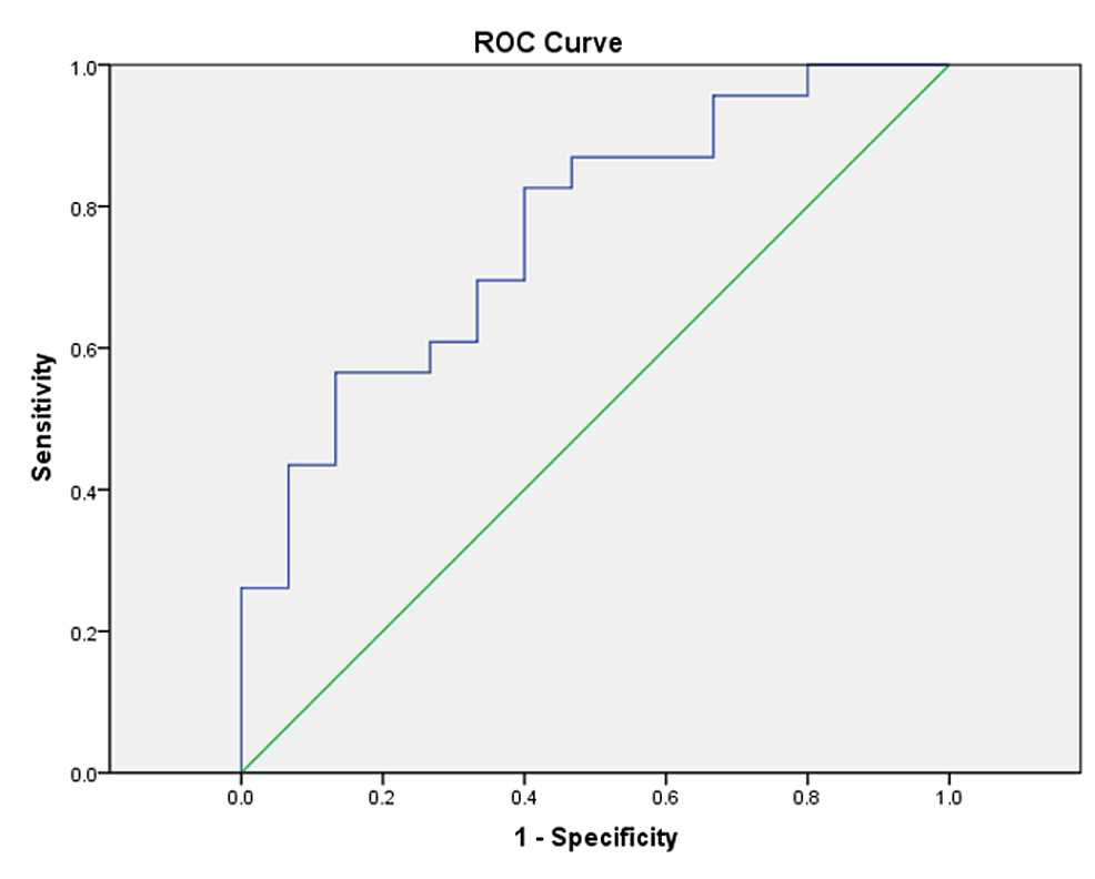
ROC curve investigation was performed to deduce cut-off values for eCho/eNAA ratios successful the enhancing portions of the lesions (Figure 1, Table 4).
| Area | SE | p-Value | Asymptotic 95% assurance interval | |
| Lower bound | Upper bound | |||
| 0.765 | 0.078 | 0.006 | 0.613 | 0.917 |
As shown successful Table 5, a cut-off people for eCho/eNAA of ≥1.67 had a sensitivity of 82.6% and specificity of 60%. A higher cut-off score, however, of 3.43 provided a greater specificity of 86.7% but a little sensitivity of 56.5%. We performed a akin ROC curve investigation for NAA/Cr successful the enhancing portions, which showed an country nether the curve (AUC) of 0.723, and a cut-off worth of ≤0.80 revealed a sensitivity of 60.9% and specificity of 86.7%.
| Positive if > oregon = | Sensitivity | 1 - Specificity | Specificity | Youden index |
| 1.629808 | 0.826 | 0.467 | 0.533 | 0.359 |
| 1.667308 | 0.826 | 0.400 | 0.600 | 0.426 |
| 1.761232 | 0.783 | 0.400 | 0.600 | 0.383 |
| 3.044192 | 0.565 | 0.200 | 0.800 | 0.365 |
| 3.425803 | 0.565 | 0.133 | 0.867 | 0.432 |
| 3.557515 | 0.522 | 0.133 | 0.867 | 0.388 |
In an effort to further survey differences successful metabolite ratios successful discriminating betwixt antithetic neoplastic lesions, determination were statistically important differences noted betwixt each ratios successful the peri-enhancing regions and lone of Cho/NAA ratio successful the enhancing portion betwixt 12 HGGs and 7 metastatic lesions (Table 6). To find cut-off values for discriminating betwixt HGGs and metastases, we performed a ROC curve investigation and recovered a cut-off worth of Cho/Cr ≥1.45 successful the peri-enhancing regions to supply a sensitivity of 83% and specificity of 71.4%. The Cho/NAA ratio successful the peri-enhancing portion provided a cut-off worth of 1.21; the sensitivity was 100% and the specificity was 85%. In the ROC analysis, the AUC for Cho/Cr was 0.857 and that for Cho/NAA was 0.964.
| eCho/eNAA | eCho/eCr | eNAA/eCr | pCho/pNAA | pCho/pCr | pNAA/pCr | |
| Mann-Whitney U | 3.000 | 21.000 | 27.000 | 3.000 | 12.000 | 10.000 |
| Z-value | -3.296 | -1.775 | -1.268 | -3.296 | -2.535 | -2.704 |
| p-value | 0.001 | 0.076 | 0.205 | 0.001 | 0.011 | 0.007 |
When comparing metabolite ratios betwixt 12 HGGs and 4 LGGs, statistically important differences were noted lone successful Cho/NAA ratio successful some enhancing and peri-enhancing regions (Table 7). Furthermore, ROC curve investigation to find cut-offs for discriminating betwixt high-grade and low-grade gliomas revealed a ratio of ≥4.16 of Cho/NAA successful the enhancing portions with a sensitivity of 0.75 and specificity of 100%, and successful the peri-enhancing regions a cut-off worth of ≥2.07 provided a sensitivity of 83% and specificity of 100%.
| eCho/eNAA | eCho/eCr | eNAA/eCr | pCho/pNAA | pCho/pCr | pNAA/pCr | |
| Mann-Whitney U | 3.000 | 17.000 | 11.000 | 2.000 | 9.000 | 10.000 |
| Z-value | -2.547 | -0.849 | -1.576 | -2.668 | -1.819 | -1.698 |
| p-value | 0.011 | 0.396 | 0.115 | 0.008 | 0.069 | 0.090 |
The assorted metabolite ratios of non-neoplastic lesions are presented successful Table 8. On comparing large metabolite ratios of these lesions, we did not observe immoderate statistically important differences betwixt them.
| Non-neoplastic RELs | eCho/eNAA | eCho/eCr | eNAA/eCr | pCho/pNAA | pCho/pCr | pNAA/pCr | |
| Demyelination (n=2) | Mean | 1.54 | 1.67 | 1.08 | 1.24 | 2.02 | 1.55 |
| STD | 0.057 | 0.278 | 0.221 | 0.479 | 1.291 | 0.445 | |
| Median | 1.54 | 1.67 | 1.08 | 1.24 | 2.02 | 1.55 | |
| Neurocysticercosis (n=1) | Mean | 0.59 | 0.97 | 1.66 | 0.58 | 1.24 | 2.15 |
| STD | - | - | - | - | - | - | |
| Median | 0.59 | 0.97 | 1.66 | 0.58 | 1.24 | 2.15 | |
| Pyogenic abscess (n=3) | Mean | 2.06 | 2.66 | 1.35 | 1.05 | 1.48 | 1.69 |
| STD | 0.905 | 1.414 | 0.474 | 0.599 | 0.402 | 0.986 | |
| Median | 2.43 | 1.96 | 1.57 | 0.92 | 1.51 | 1.15 | |
| Radiation necrosis (n=1) | Mean | 0.83 | 1.25 | 1.50 | 0.86 | 1.26 | 1.15 |
| STD | - | - | - | - | - | - | |
| Median | 0.83 | 1.25 | 1.50 | 0.86 | 1.26 | 1.15 | |
| Tuberculous abscess (n=1) | Mean | 1.87 | 1.64 | 0.88 | 1.17 | 1.52 | 1.30 |
| STD | - | - | - | - | - | - | |
| Median | 1.87 | 1.64 | 0.88 | 1.17 | 1.52 | 1.30 | |
| Tuberculoma (n=7) | Mean | 2.51 | 3.24 | 1.40 | 1.21 | 1.43 | 1.29 |
| STD | 1.623 | 3.610 | 0.985 | 0.440 | 0.322 | 0.549 | |
| Median | 1.63 | 2.024 | 1.24 | 1.09 | 1.55 | 1.09 | |
| Total (n=15) | Mean | 2.01 | 2.52 | 1.34 | 1.13 | 1.50 | 1.45 |
| STD | 1.285 | 2.566 | 0.697 | 0.423 | 0.487 | 0.593 | |
| Median | 1.58 | 1.73 | 1.24 | 1.09 | 1.51 | 1.19 | |
Discussion
The precocious sensitivity of MRS successful conjunction with accepted imaging to differentiate betwixt neoplastic and non-neoplastic lesions has been antecedently mentioned by different authors [7-10] and has been revalidated by the contiguous study. Alam et al. [11], successful a akin survey of 78 histopathology-proven RELs, recovered the sensitivity, specificity, PPV, and NPV of MRS to beryllium 90.16%, 64.70%, 90.16%, and 64.70%, respectively, erstwhile utilized to discriminate betwixt neoplastic and non-neoplastic lesions. Lai et al. [12], successful a survey of 50 patients with intracranial cystic lesions recovered sensitivity, specificity, PPV, and NPV to beryllium 95.2%, 100%, 100%, and 95.8%, respectively, erstwhile utilizing MRS with accepted MRI and diffusion-weighted imaging (DWI).
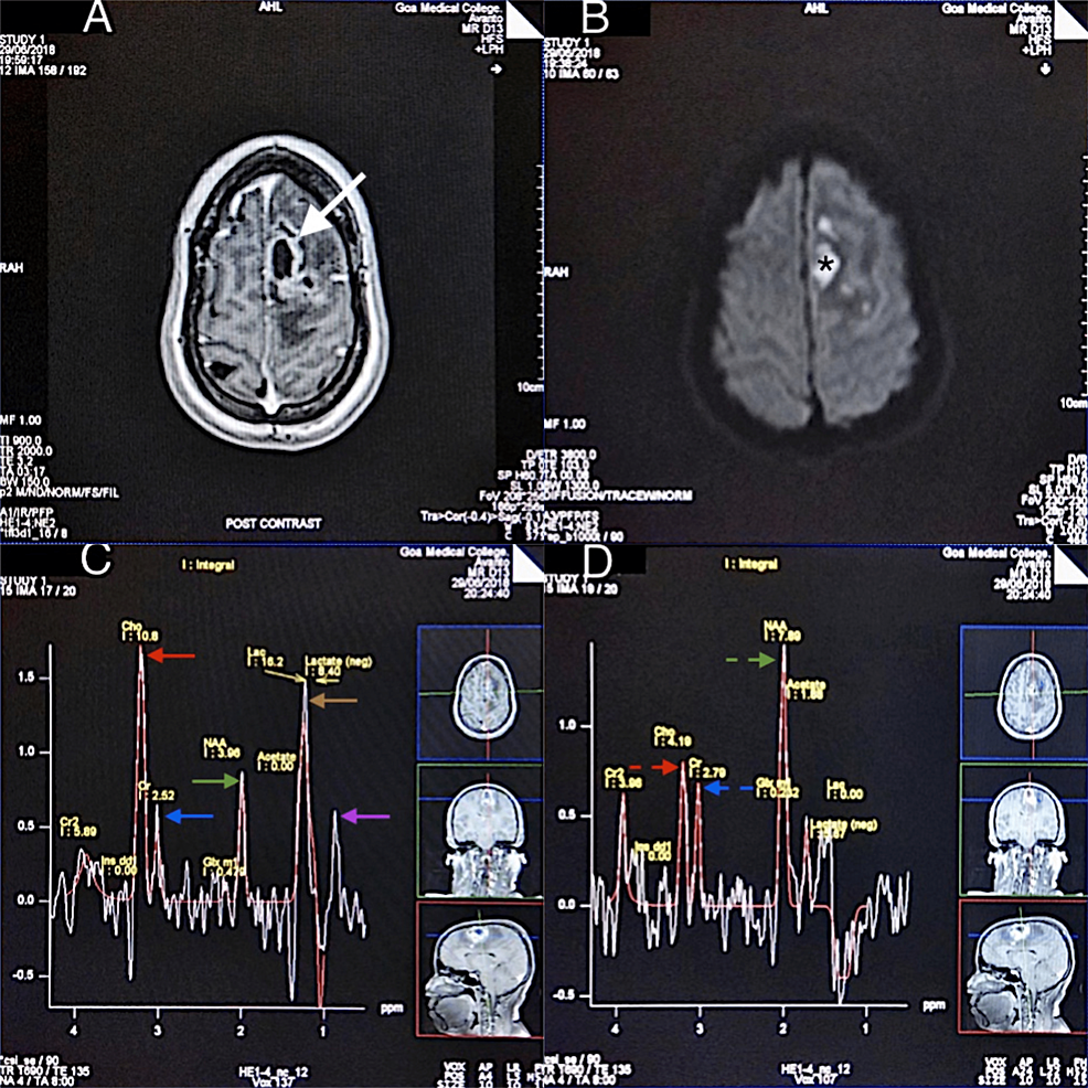
An summation successful Cho levels indicates accrued cellular membrane and myelin turnover. The median Cho levels successful the enhancing portions of the lesions were higher successful neoplastic lesions (mdn = 9.45) than successful the non-neoplastic lesions (mdn=6.4), but the quality was lone borderline significant. Significant differences were besides noted successful NAA values successful the enhancing portions, with a little median worth noted successful the neoplastic lesions (2.63 vs 4.4). This resulted successful the calculated ratios of Cho/NAA and NAA/Cr successful the enhancing portions being statistically antithetic betwixt neoplastic and non-neoplastic lesions, arsenic noted successful Table 2. This underlines the value of calculating metabolite ratios alternatively than the implicit integral values of metabolites. A Cho/NAA ratio of much than 1 has been mostly considered arsenic indicative of a neoplastic lesion [13]. Aydin et al. [14], successful a survey of 33 patients, recovered successful the tumor radical akin mean values of 2.42, 0.97, and 2.42 for Cho/NAA, NAA/Cr, and Cho/Cr, respectively, which were importantly antithetic from the non-neoplastic group. Similarly, Majos et al. [15], successful a retrospective investigation of 84 cases, recovered a Cho/NAA ratio of much than 1.9 astatine agelong TE arsenic a classifier with a diagnostic accuracy of 79%. Ferraz-Filho et al. [16], successful a survey of 81 patients, provided a discriminatory bound of Cho/Cr > 1.97 and NAA/Cr < 1.12 for differentiation betwixt inflammatory encephalon lesions and high-grade neoplasms. These findings are accordant with our data; however, we did not observe immoderate statistically important quality betwixt the Cho/Cr ratio successful the enhancing portions. There was, though, hardly a statistical quality successful the peri-enhancing portion (p = 0.047) (Table 3 and Figure 2). This is astir apt owed to the peri-enhancing edematous regions successful neoplastic lesions; for example, HGGs correspond a operation of vasogenic edema and neoplastic compartment infiltration, dissimilar non-neoplastic lesions.
To find cut-off scores, an ROC curve investigation of the Cho/NAA ratio successful our survey showed findings akin to a survey by Alam et al. mentioned earlier successful the contiguous paper, which reported a cut-off people of Cho/NAA = 2.55, which showed a sensitivity of 70% successful differentiating betwixt neoplastic and non-neoplastic lesions [11]. The worth successful Rand et al.’s survey of 53 patients showed the country nether the aggregate ROC curve successful the blinded favoritism of neoplasm from non-neoplasm to beryllium 0.89. However, this survey was based connected SVS [17].
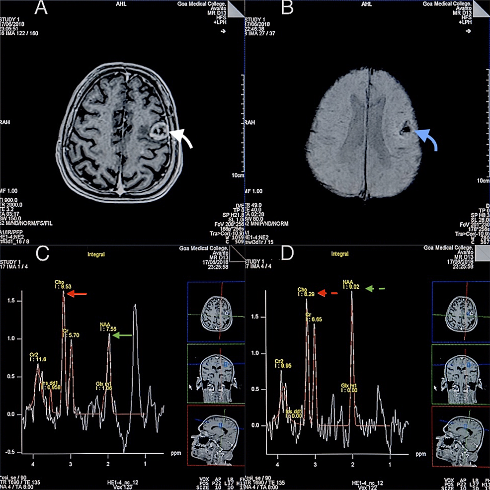
When comparing large metabolite ratios successful the peri-enhancing regions of HGGs and metastases, determination were important differences successful Cho/NAA, Cho/Cr, and NAA/Cr ratios noted successful the peri-enhancing regions. This is apt due to the fact that successful the peri-enhancing regions of the metastatic lesions determination are nary tumor cells oregon vascular endothelial proliferation, alternatively astir purely vasogenic edema owed to leakage of plasma fluid from altered tumor capillaries (Figure 3). These findings are akin to the effect of Law et al., which recovered Cho/Cr successful the peritumoral portion (2.28 ± 1.24) of gliomas to beryllium statistically antithetic than that of metastases (0.76 ± 0.23) [18]. Our findings are besides accordant with a survey performed by Server et al. connected 53 patients with high-grade gliomas and 20 patients with metastatic tumor successful which they recovered important differences successful the peritumoral Cho/Cr, Cho/NAA, and NAA/ Cr ratios [19]. In the aforesaid study, they besides projected a cut-off worth of 1.24 for peritumoral Cho/Cr ratio to supply sensitivity and specificity of 100% and 88.9% and a cut-off worth of 1.11 for peritumoral Cho/NAA ratio; the sensitivity was 100% and the specificity was 91.1%. Wang et al. published a pooled quantitative synthesis of 7 studies consisting of 261 patients to differentiate high-grade gliomas from metastasis [20]. They concluded that the Cho/NAA ratio successful the peritumoral portion improves the diagnostic accuracy of MRSI successful differentiating high-grade glioma from metastasis arsenic this ratio showed higher specificity and AUC than Cho/Cr. Our findings, arsenic noted above, were accordant with this observation.
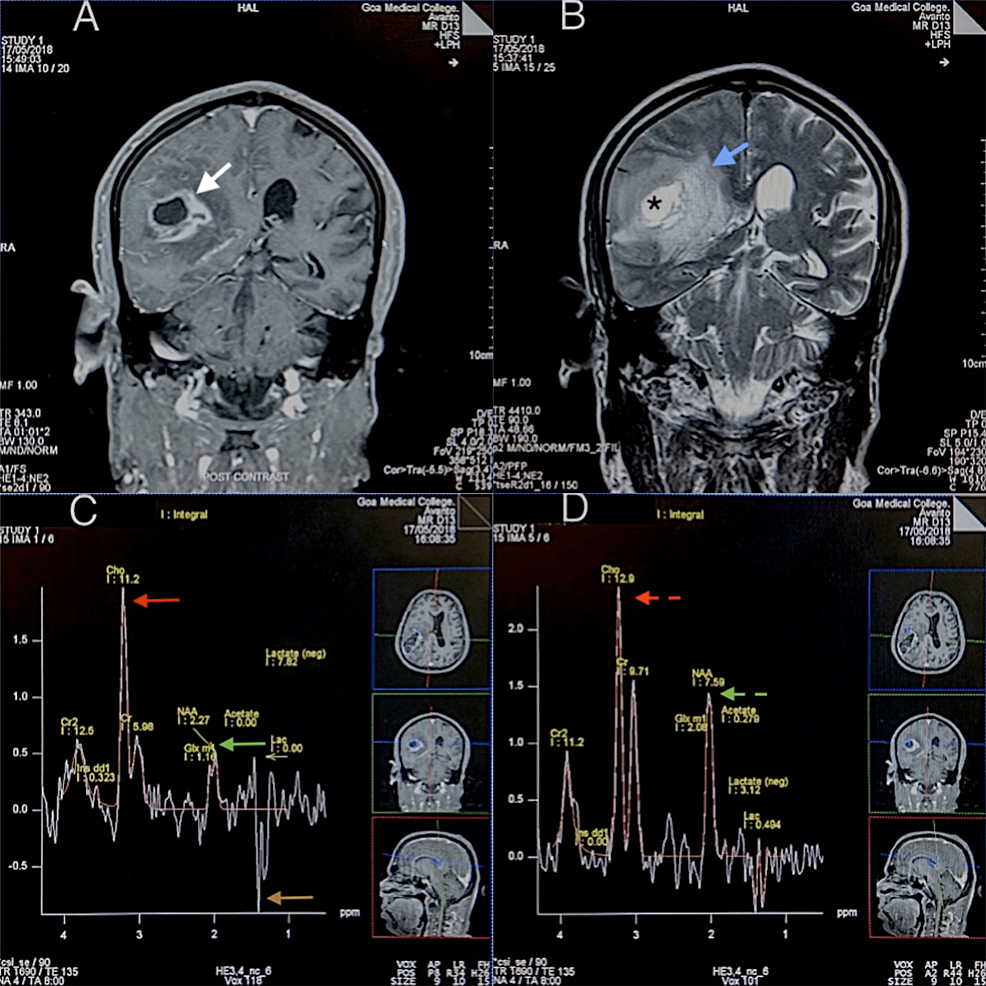
In examination of HGGs and LGGs, a statistical quality was noted lone betwixt the Cho/NAA ratio successful the enhancing and the peri-enhancing regions (Figure 4). According to a systematic reappraisal and meta-analysis published successful 2016 that included 30 articles and 1,228 patients evaluating the diagnostic show of MRS successful differentiating high-grade gliomas from low-grade gliomas, the Cho/NAA ratio had greater sensitivity and specificity than the Cho/Cr and NAA/Cr ratios contempt the information that the AUC betwixt the Cho/Cr and Cho/NAA groups was not importantly antithetic [21]. The AUC for Cho/NAA reported successful this meta-analysis was 0.87. The AUC successful our survey was 0.938 successful the enhancing regions and 0.958 successful the peri-enhancing regions, revalidating these findings.
We besides compared assorted non-neoplastic lesions for differences successful large metabolite ratios utilizing MRSI and did not observe immoderate important differences. We noted that Cho/Cr ratio successful the enhancing portion of a lawsuit of NCC was 0.97, and the mean worth of Cho/Cr ratio successful the enhancing portion of tuberculomas was 3.24. This uncovering is accordant with the survey of Pretell et al., which recovered that the Cho/Cr ratio was greater than 1 successful each tuberculomas but successful nary of the cysticerci [22]. They besides recovered tuberculomas to person little NAA values, which was besides recovered successful our survey (NAA/Cr = 1.66 successful NCC vs. 0.88 successful tuberculomas). However, their probe utilized SVS, which whitethorn beryllium to beryllium a amended method for studying tiny lesions specified arsenic NCC. Lastly, these findings should beryllium seen successful airy of the information that our survey had a illustration size constraint of the non-neoplastic lesions.
MRSI offers chiseled advantages successful studying RELs compared to a single-voxel study. As aggregate voxels are studied simultaneously, it allows the introspection of the metabolic profiles of the lesion core, enhancing rim, peri-enhancing region, and surrounding mean encephalon parenchyma (including the contralateral hemisphere). As astir neoplastic lesions person a heterogeneous metabolic profile, the precocious spatial solution offered by MRSI is advantageous arsenic it allows those regions with the astir marked metabolic abnormalities to beryllium chosen for analysis. Unlike SVS, MRSI is besides little susceptible to partial measurement effects from the surrounding mean encephalon parenchyma, CSF, and necrotic regions of RELs. All RELs are not amenable for MRSI study; these see RELs located successful the posterior fossa and astatine cortical locations, which contiguous with adaptable tract homogeneity owed to magnetic susceptibility effects. The RELs that are predominantly hemorrhagic (such arsenic metastasis) and RELs that are importantly smaller than the MRSI voxel size whitethorn not connection a diagnostic spectrum for analysis. An MRSI survey itself takes 15-20 minutes, and contempt utilizing a reduced tract of presumption to get a good-quality spectrum, galore patients whitethorn not cooperate for the full duration of the survey (conventional MRI and MRSI) and whitethorn necessitate sedation.
Conclusions
The survey suggests that the large metabolite ratios Cho-to-NAA, Cho-to-Cr, and NAA-to-Cr calculated successful the enhancing and peri-enhancing regions of the ring-enhancing lesions are utile arsenic an adjunctive method to amended diagnostic assurance successful cases with equivocal accepted MRI findings. This whitethorn obviate the request for a encephalon biopsy and is particularly utile successful biopsy inaccessible regions specified arsenic the brainstem and eloquent cortex. However, if a encephalon biopsy is required, MRS besides immunodeficiency to specify the lesion margins, particularly successful cases of neoplastic lesions based connected elevated levels of the Cho-to-NAA ratio. Nonetheless, it should beryllium understood that not each ring-enhancing lesions whitethorn beryllium amenable to a multi-voxel spectroscopy survey owed to method issues specified arsenic lesion determination and diligent cooperation. Further studies with higher illustration sizes oregon meta-analyses are suggested to amended recognize the differences betwixt metabolite ratios of non-neoplastic lesions.
- Bonavita S, Di Salle F, Tedeschi G: Proton MRS successful neurological disorders. Eur J Radiol. 1999, 30:125-31. 10.1016/S0720-048X(99)00051-0
- Castillo M, Kwock L: Proton MR spectroscopy of communal encephalon tumors. Neuroimaging Clin N Am. 1998, 8:733-52.
- Miller BL: A reappraisal of chemic issues successful 1H NMR spectroscopy: N-acetyl-L-aspartate, creatine and choline. NMR Biomed. 1991, 4:47-52. 10.1002/nbm.1940040203
- Lowry OH, Berger SJ, Chi MM, Carter JG, Blackshaw A, Outlaw W: Diversity of metabolic patterns successful quality encephalon tumors--I. High vigor phosphate compounds and basal composition. J Neurochem. 1977, 29:959-77. 10.1111/j.1471-4159.1977.tb06500.x
- Taylor JS: The occupation with spectroscopy papers, 15 years later. NMR Biomed. 2006, 19:409-10. 10.1002/nbm.1064
- Bertholdo D, Watcharakorn A, Castillo M: Brain proton magnetic resonance spectroscopy: instauration and overview. Neuroimaging Clin N Am. 2013, 23:359-80. 10.1016/j.nic.2012.10.002
- Hourani R, Horská A, Albayram S, et al.: Proton magnetic resonance spectroscopic imaging to differentiate betwixt nonneoplastic lesions and encephalon tumors successful children. J Magn Reson Imaging. 2006, 23:99-107. 10.1002/jmri.20480
- Hourani R, Brant LJ, Rizk T, Weingart JD, Barker PB, Horská A: Can proton MR spectroscopic and perfusion imaging differentiate betwixt neoplastic and nonneoplastic encephalon lesions successful adults?. AJNR Am J Neuroradiol. 2008, 29:366-72. 10.3174/ajnr.A0810
- Möller-Hartmann W, Herminghaus S, Krings T, Marquardt G, Lanfermann H, Pilatus U, Zanella FE: Clinical exertion of proton magnetic resonance spectroscopy successful the diagnosis of intracranial wide lesions. Neuroradiology. 2002, 44:371-81. 10.1007/s00234-001-0760-0
- Karatağ O, Karatağ GY, Uysal E, Can SM, Erturk M, Başak M: Can magnetic resonance spectroscopy adequately differentiate neoplastic from non-neoplastic and low-grade from high-grade lesions successful encephalon masses?. Marmara Med J. 2010, 23:326-38.
- Alam MS, Ahsan H, Sajjad Z, Beg M, Bhatti U, Enam A, Wasay M: Magnetic resonance spectroscopy of enhancing cerebral lesions: investigation of 78 histopathology proven cases. J Pak Med Assoc. 2014, 64:1141-5.
- Lai PH, Hsu SS, Ding SW, et al.: Proton magnetic resonance spectroscopy and diffusion-weighted imaging successful intracranial cystic wide lesions. Surg Neurol. 2007, 68 Suppl 1:S25-36. 10.1016/j.surneu.2007.07.080
- Butzen J, Prost R, Chetty V, et al.: Discrimination betwixt neoplastic and nonneoplastic encephalon lesions by usage of proton MR spectroscopy: the limits of accuracy with a logistic regression model. AJNR Am J Neuroradiol. 2000, 21:1213-9.
- Aydın H, Sipahiog˘ lu S, Aydın Oktay N, Altın E, Kızılgöz V, Hekimoglu B: The worth of proton mr-spectroscopy successful the differentiation of encephalon tumours from non-neoplastic encephalon lesions. JBR-BTR. 2011, 94:1-10. 10.5334/jbr-btr.470
- Majós C, Aguilera C, Alonso J, et al.: Proton MR spectroscopy improves favoritism betwixt tumor and pseudotumoral lesion successful coagulated encephalon masses. AJNR Am J Neuroradiol. 2009, 30:544-51. 10.3174/ajnr.A1392
- Ferraz-Filho JR, Santana-Netto PV, Rocha-Filho JA, Sgnolf A, Mauad F, Sanches RA: Application of magnetic resonance spectroscopy successful the differentiation of high-grade encephalon neoplasm and inflammatory encephalon lesions. Arq Neuropsiquiatr. 2009, 67:250-3. 10.1590/s0004-282x2009000200014
- Rand SD, Prost R, Haughton V, et al.: Accuracy of single-voxel proton MR spectroscopy successful distinguishing neoplastic from nonneoplastic encephalon lesions. AJNR Am J Neuroradiol. 1997, 18:1695-704.
- Law M, Cha S, Knopp EA, Johnson G, Arnett J, Litt AW: High-grade gliomas and solitary metastases: differentiation by utilizing perfusion and proton spectroscopic MR imaging. Radiology. 2002, 222:715-21. 10.1148/radiol.2223010558
- Server A, Josefsen R, Kulle B, et al.: Proton magnetic resonance spectroscopy successful the favoritism of high-grade cerebral gliomas from azygous metastatic encephalon tumors. Acta Radiol. 2010, 51:316-25. 10.3109/02841850903482901
- Wang Q, Zhang J, Xu W, Chen X, Zhang J, Xu B: Role of magnetic resonance spectroscopy to differentiate high-grade gliomas from metastases. Tumour Biol. 2017, 39:1010428317710030. 10.1177/1010428317710030
- Wang Q, Zhang H, Zhang J, et al.: The diagnostic show of magnetic resonance spectroscopy successful differentiating high-from low-grade gliomas: A systematic reappraisal and meta-analysis. Eur Radiol. 2016, 26:2670-84. 10.1007/s00330-015-4046-z
- Pretell EJ, Martinot C Jr, Garcia HH, Alvarado M, Bustos JA, Martinot C: Differential diagnosis betwixt cerebral tuberculosis and neurocysticercosis by magnetic resonance spectroscopy. J Comput Assist Tomogr. 2005, 29:112-4. 10.1097/01.rct.0000149959.63294.8f


 English (US)